Uploads by Shamilton
This special page shows all uploaded files.
| Date | Name | Thumbnail | Size | Description | Versions |
|---|---|---|---|---|---|
| 14:19, 4 August 2008 | TheSpinocerebellarTract.jpg (file) |  |
8 KB | 1 | |
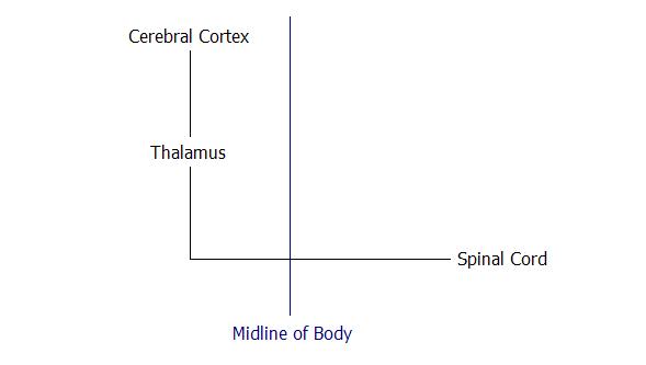
| 11:51, 4 August 2008 | The Ascending Pathways.jpg (file) |  |
9 KB | 1 | |
| 11:46, 4 August 2008 | AscendingReticularFormation.jpg (file) |  |
11 KB | 1 | |
| 11:44, 4 August 2008 | SpinocerebellarTract.jpg (file) |  |
8 KB | 1 | |
| 11:40, 4 August 2008 | AscendingPathways.jpg (file) |  |
9 KB | 1 | |
| 11:38, 4 August 2008 | Pyramidalsystem.jpg (file) |  |
9 KB | 1 | |
| 11:03, 4 August 2008 | Extrapyramidal system.jpg (file) |  |
17 KB | 1 | |
| 15:06, 1 August 2008 | Pyramidal system.jpg (file) |  |
9 KB | 1 | |
| 15:02, 1 August 2008 | Pyramidal System.jpg (file) |  |
9 KB | 1 | |
| 12:45, 31 July 2008 | Ascending Reticular Formation.jpg (file) |  |
11 KB | 1 | |
| 10:38, 31 July 2008 | Spinocerebellar Tract.jpg (file) |  |
8 KB | 1 | |
| 10:29, 31 July 2008 | Ascending Pathways.jpg (file) |  |
9 KB | 1 | |
| 07:24, 31 July 2008 | Sarah.jpg (file) |  |
17 KB | 1 | |
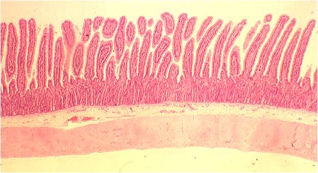
| 15:12, 28 July 2008 | Tunica muscularis.jpg (file) |  |
119 KB | 1 | |
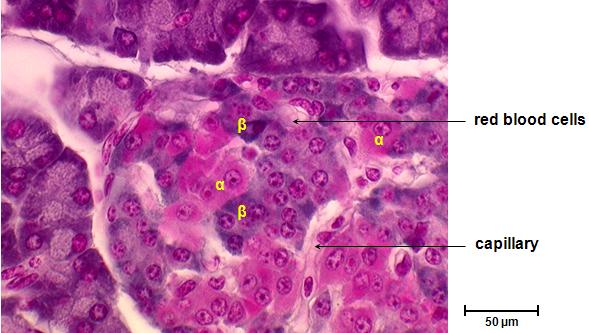
| 08:31, 22 July 2008 | Islet cell types.jpg (file) |  |
38 KB | 1 | |
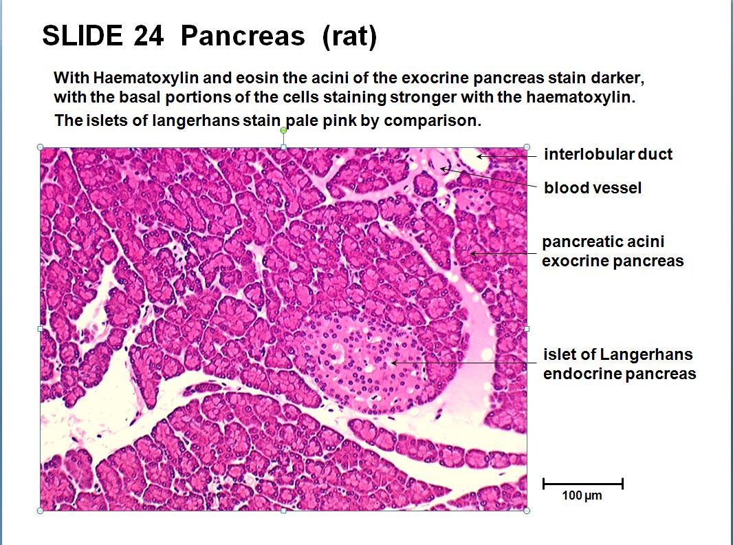
| 15:06, 21 July 2008 | Islet and acini.jpg (file) |  |
174 KB | 1 | |
| 15:03, 21 July 2008 | Acini and islet.jpg (file) |  |
179 KB | 1 | |
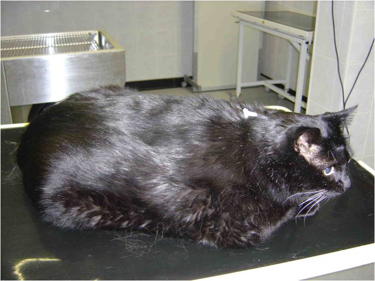
| 11:03, 18 July 2008 | NIDDM cat.jpg (file) |  |
57 KB | 1 | |
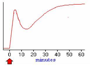
| 10:00, 18 July 2008 | Insulin Secretion Pattern.jpg (file) |  |
7 KB | 1 | |
| 09:40, 18 July 2008 | Pancreatic Beta Cell.jpg (file) |  |
32 KB | 1 | |
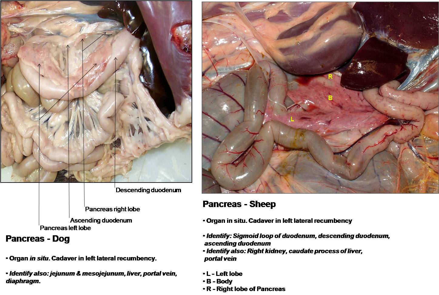
| 14:32, 17 July 2008 | Sheep Pancreas.jpg (file) |  |
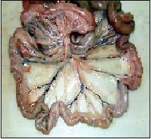
70 KB | R=Right lobe B=Body L=Left lobe | 1 |
| 14:12, 17 July 2008 | Pancreas Sheep.jpg (file) |  |
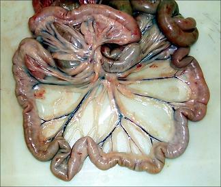
195 KB | The pancreas can be seen dorsally to the descending duodenum. A left and right lobe and body can be seen. | 1 |
| 10:30, 17 July 2008 | Recto-Anal Junction.jpg (file) |  |
84 KB | 1 | |
| 07:43, 17 July 2008 | Illustration dog descending colon.jpg (file) |  |
38 KB | 1 | |
| 07:40, 17 July 2008 | Opendogcaecum.jpg (file) |  |
51 KB | 1 | |
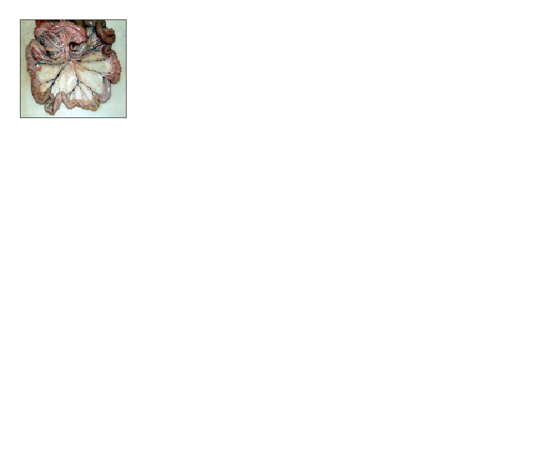
| 07:35, 17 July 2008 | Small&largeintestine.jpg (file) |  |
53 KB | 1 | |
| 07:33, 17 July 2008 | Smalllargeintestine.jpg (file) |  |
53 KB | 1 | |
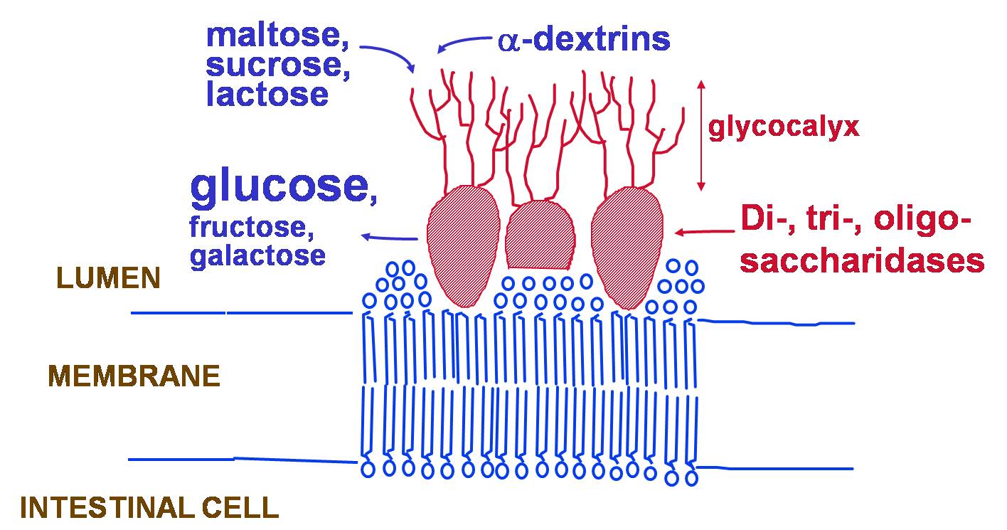
| 10:09, 16 July 2008 | Dissacharidase.jpg (file) |  |
136 KB | 1 | |
| 10:21, 15 July 2008 | Cellulose.jpg (file) |  |
48 KB | 1 | |
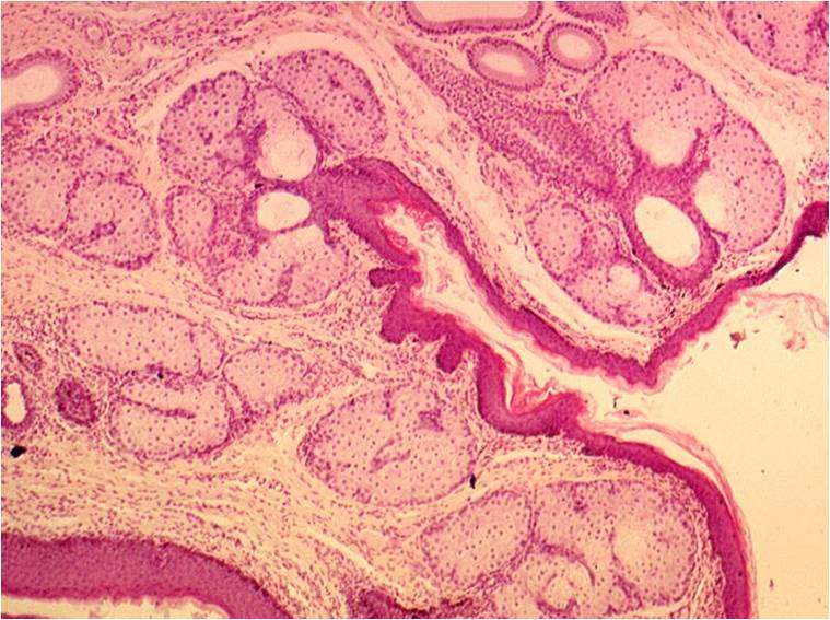
| 14:49, 14 July 2008 | Anal Glands.jpg (file) |  |
91 KB | Anal glands in the lamina propria of the mucosa. | 1 |
| 14:47, 14 July 2008 | Anal Sacs.jpg (file) |  |
91 KB | Anal glands beneath the epithelium | 1 |
| 14:00, 14 July 2008 | Mucosal layer of colon.jpg (file) |  |
107 KB | 1 | |
| 13:55, 14 July 2008 | Colon fox lymphatic nodule.jpg (file) |  |
109 KB | A lymphatic nodule from the colon of the fox | 1 |
| 13:50, 14 July 2008 | Colon fox transverse section.jpg (file) | 80 KB | A transverse section through the colon of the fox | 1 | |
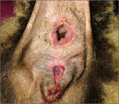
| 13:46, 14 July 2008 | Sheep Anus.jpg (file) |  |
47 KB | 1 | |
| 15:24, 7 July 2008 | Caudalduodenum.jpg (file) |  |
35 KB | 1 | |
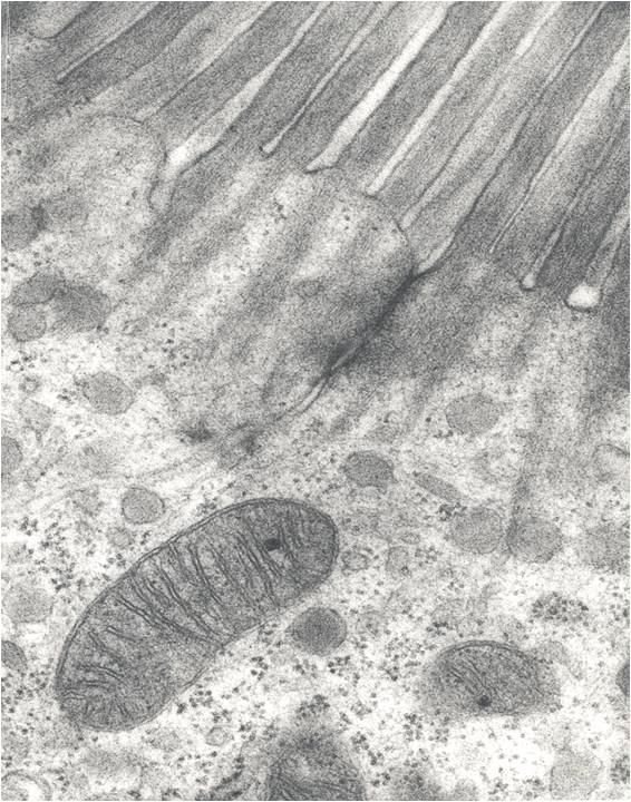
| 15:18, 7 July 2008 | Electronmicrographduodenum.jpg (file) |  |
99 KB | 1 | |
| 19:07, 5 July 2008 | Jejunum1.jpg (file) |  |
22 KB | 1 | |
| 19:06, 5 July 2008 | Intestinesjejunum.jpg (file) |  |
22 KB | 1 | |
| 16:32, 5 July 2008 | Jejunum.jpg (file) |  |
13 KB | 1 | |
| 16:31, 5 July 2008 | Jejunumphoto2.jpg (file) |  |
28 KB | 1 | |
| 16:30, 5 July 2008 | Jejunumphoto1.jpg (file) |  |
28 KB | 1 | |
| 16:26, 5 July 2008 | Jejunumphoto.jpg (file) |  |
138 KB | 1 |