Unused files
Jump to navigation
Jump to search
The following files exist but are not embedded in any page. Please note that other web sites may link to a file with a direct URL, and so may still be listed here despite being in active use.
Showing below up to 250 results in range #21 to #270.
View (previous 250 | next 250) (20 | 50 | 100 | 250 | 500)
 Kim.jpg 200 × 299; 9 KB
 IMG 1366.jpg 800 × 497; 595 KB
 Alex.jpg 200 × 311; 10 KB
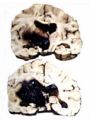
 Cowhead.jpg 233 × 350; 46 KB
 Picture.jpg 683 × 471; 98 KB
 Cat.jpg 330 × 191; 21 KB
 Test.ppt ; 619 KB
 COP 310707.JPG 800 × 347; 201 KB
 Funny cats.jpg 640 × 480; 91 KB
 Pteridium aquilinum plant.jpg 593 × 600; 172 KB
 Cleft-palate.gif 683 × 442; 208 KB
 Uraemia.gif 673 × 442; 170 KB
 BVD-MD.gif 723 × 396; 227 KB
 Bvd2.gif 586 × 382; 188 KB
 BPS.gif 781 × 426; 208 KB
 BPSOES.gif 750 × 373; 168 KB
 MCF1.gif 620 × 402; 121 KB
 MCF2.gif 483 × 327; 116 KB
 Woodentongue1.gif 433 × 682; 189 KB
 Woodentongue2.gif 434 × 313; 121 KB
 Lumpyjaw1.gif 594 × 396; 150 KB
 Dogpap1.gif 337 × 512; 126 KB
 Cowpap1.gif 736 × 505; 202 KB
 Fibrosarc.gif 610 × 394; 164 KB
 Epulis.gif 631 × 414; 177 KB
 Tonguelymphoma.gif 237 × 649; 103 KB
 Enamal-hypoplasia.gif 833 × 553; 246 KB
 Oligodontia.gif 334 × 500; 127 KB
 Toothinfection.gif 662 × 480; 201 KB
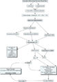
 CmapTools - Home Page Cmap.png 2,792 × 1,922; 390 KB
 CmapTools - Home Page Cmap.pdf ; 280 KB
 Praa.gif 706 × 450; 237 KB
 Megaoes.gif 678 × 438; 215 KB
 Bovimpaction.gif 343 × 292; 60 KB
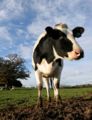 Cow2.jpg 300 × 392; 24 KB
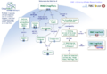
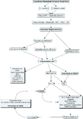
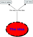 Mindmap.png 774 × 874; 41 KB
- Mindmap.pdf ; 23 KB
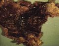 Haemorrhagic gastritis.jpg 742 × 572; 63 KB
 Abomasal lymphoma.jpg 818 × 556; 68 KB
 Adenocarcinoma stomach histopath2.jpg 738 × 574; 64 KB
 Adenocarcinoma stomach.jpg 742 × 576; 69 KB
 Oesophageal bloat line.jpg 811 × 526; 58 KB
 Traumatic pericarditis.jpg 320 × 256; 7 KB
 Gastric ulcer.jpg 738 × 576; 65 KB
 Gastric ulcer histopath.jpg 746 × 540; 55 KB
 Ostertagiasis.jpg 762 × 524; 57 KB
 Leiomyoma.jpg 742 × 572; 67 KB
 Acute interstitial pancreatitis.jpeg 467 × 302; 30 KB
 Chronic pancreatitis.jpeg 523 × 332; 41 KB
 Normal perianal gland.jpg 746 × 570; 97 KB
 Adenoma.jpeg 402 × 652; 83 KB
 Perianal gland adenoma histopath.jpg 750 × 576; 100 KB
 Perianal gland adenoma.jpg 626 × 570; 51 KB
 Strongylus vulgaris.jpg 760 × 548; 78 KB
 Adenoma2.jpeg 652 × 400; 82 KB
 Carcinoma.jpeg 533 × 357; 30 KB
 Carcinoma gross.jpeg 533 × 357; 30 KB
 Carcinoma micro.jpeg 469 × 312; 44 KB
 Infaction of the small bowel.jpg 744 × 576; 65 KB
 Pancreatic nodular hyperplasia.jpeg 510 × 403; 36 KB
 Islet.jpeg 466 × 270; 48 KB
 Pancreatic hypoplasia.jpeg 229 × 199; 10 KB
 Pancreatic hypoplasia micro.jpeg 445 × 287; 34 KB
 Brunner gland adenoma.jpg 742 × 574; 75 KB
 Acute haemorrhagic pancreatitis.jpeg 479 × 311; 37 KB
 Acute pancreatic necrosis.jpeg 429 × 234; 30 KB
 Insulinoma.jpeg 442 × 287; 56 KB
 Beta cell carcinoma.jpeg 450 × 284; 48 KB
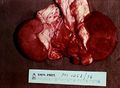
 Atresia ani PM.jpg 807 × 539; 94 KB
 Gill2.jpg 179 × 202; 14 KB
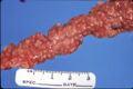
 Johnes disease proliferative enteritis.jpg 762 × 550; 50 KB
 Pulpy kidney disease.jpg 320 × 256; 15 KB
 Pulpy kidney gross.jpg 762 × 556; 70 KB
 Bucket and spade.jpg 464 × 298; 14 KB
 Trichuris vulpis caecum.jpg 762 × 552; 100 KB
 Trichuris vulpis caecum comparative.jpg 766 × 528; 69 KB
 Trichuris ovis.jpg 762 × 528; 55 KB
 Johnes disease histological.jpg 762 × 558; 100 KB
 Johnes disease proliferative ileitis.jpg 764 × 522; 63 KB
 Porcine intestinal adenomatosis campylobacter.jpg 764 × 528; 107 KB
 Intussusception.jpg 622 × 332; 34 KB
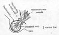 Hernial sac.jpg 713 × 424; 51 KB
 Stomach diaphragmatic hernia.jpg 320 × 256; 13 KB
 Volvulus.jpg 762 × 542; 54 KB
 Intussuceptionphoto.jpg 744 × 572; 56 KB
 Chronic peritonitis with fibrosis.jpeg 750 × 491; 74 KB
 FIP.jpeg 750 × 500; 55 KB
 Omentum carcinoma.jpeg 750 × 504; 308 KB
 Bile stained peritonitis and gastric rupture.jpeg 750 × 497; 66 KB
 FIP severe exudative peritonitis.jpeg 750 × 571; 36 KB
 Nocardiosis in a puma.jpeg 750 × 497; 310 KB
 Glasser's disease - severe acute fibrinous peritonitis.jpeg 750 × 493; 75 KB
 Acute peritonitis and cecal base rupture.jpeg 750 × 508; 100 KB
 Cysticercus pisiformis.jpeg 750 × 497; 276 KB
 Tubeculous peritonitis.jpeg 320 × 248; 13 KB
 Carcinomatosis and sclerosis in sheep.jpeg 750 × 499; 189 KB
 Lipoma in horse.jpeg 750 × 499; 63 KB
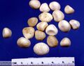 Bovine pancreatic calculi.jpeg 750 × 594; 50 KB
 Pancreatic hypoplasia by King.jpeg 750 × 513; 50 KB
 Pancreatic adenoma cat.jpeg 750 × 499; 299 KB
 Ectopic pancreas.jpeg 1,796 × 1,076; 57 KB
 Insulinoma King.jpeg 750 × 498; 197 KB
 Pancreatic cysts by.jpeg 750 × 516; 352 KB
 Pancreatic flukes in a wolf by King.jpeg 750 × 500; 177 KB
 Fat necrosis by King.jpeg 750 × 487; 99 KB
 Pancreatic carcinoma.jpeg 750 × 480; 66 KB
 Strongylus equinus granulomas in pancreas.jpeg 750 × 515; 101 KB
 Gastrinoma by King.jpeg 750 × 496; 62 KB
 Peritoneal mesothelioma BioMed by King.jpeg 320 × 247; 16 KB
 Congenital umbilical hernia.jpeg 750 × 563; 53 KB
 Rupture uterus with fibrinous peritonitis in a cow.jpeg 750 × 504; 540 KB
 Rupture pyometra in a rabbit.jpeg 750 × 500; 173 KB
 Diaphragmatic hernia in a cat in RTA.jpeg 750 × 497; 64 KB
 Steatitis.jpeg 750 × 516; 62 KB
 Granulomatous fat necrosis in Guernsey.jpeg 750 × 514; 55 KB
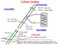 Cellular swelling diagram.jpg 710 × 543; 56 KB
 Hydropic degeneration foot and mouth pig foot.jpg 247 × 552; 23 KB
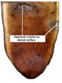 Hydropic degneration foot and mouth ox tongue.jpg 292 × 391; 25 KB
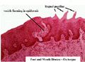 Hydropic degeneration foot and mouth ox tongue histo 1.jpg 326 × 236; 16 KB
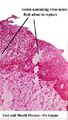 Hydropic degeneration foot and mouth ox tongue histo 2.jpg 188 × 333; 17 KB
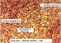
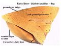 Fatty liver.jpg 404 × 306; 21 KB
 Fatty liver histo.jpg 547 × 387; 68 KB
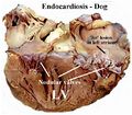 Endocardiosis.jpg 496 × 436; 45 KB
 Endocardiosis histo 2.jpg 493 × 340; 35 KB
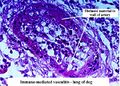 Fibrinoid degeneration immune mediated vasculitis.jpg 474 × 338; 54 KB
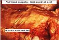 Nutritional myopathy.jpg 478 × 325; 34 KB
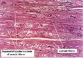 Nutritional myopathy histo.jpg 453 × 318; 39 KB
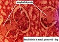 Amyloidosis.jpg 411 × 293; 29 KB
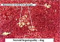 Glycogen infiltration.jpg 496 × 350; 53 KB
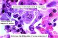 Viral inclusion canine adenovirus 1.jpg 363 × 237; 23 KB
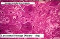 Lysosomal storage disease.jpg 526 × 347; 41 KB
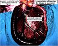 Strangulation of intestine.jpg 512 × 404; 49 KB
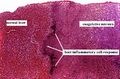
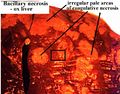 Coagulative necrosis bacillary necrosis.jpg 437 × 343; 35 KB
 Coagulative necrosis histo.jpg 422 × 277; 35 KB
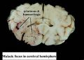 Malacia.jpg 448 × 320; 19 KB
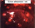 Liver abscess.jpg 379 × 304; 21 KB
 Abscess slice.jpg 506 × 240; 31 KB
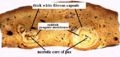
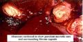 Abscess centre and capsule.jpg 368 × 186; 19 KB
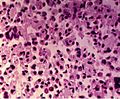 Macrophage caseative necrosis.JPG 421 × 348; 39 KB
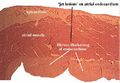
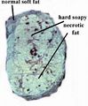 Fat necrosis ox subcutis.jpg 294 × 356; 18 KB
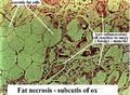 Fat necrosis ox subcutis histo.jpg 363 × 266; 33 KB
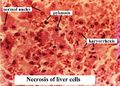 Necrosis histo.jpg 465 × 334; 37 KB
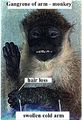 Monkey gangrene 1.jpg 337 × 495; 41 KB
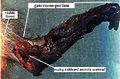 Monkey gangrene 2.jpg 395 × 259; 27 KB
 Calcification.jpg 320 × 210; 19 KB
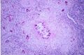
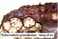 Tuberculosis granuloma.jpg 421 × 288; 29 KB
 Nick Photo.jpg 312 × 302; 16 KB
 Nick Short.jpg 312 × 302; 16 KB
 Clot.jpg 280 × 220; 8 KB
 Hypostatic congestion.jpg 264 × 208; 11 KB
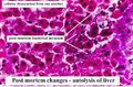 Autolysis of liver.jpg 301 × 197; 21 KB
 Pseudomelanosis.jpg 365 × 261; 24 KB
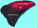
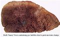 Foamy liver.jpg 461 × 283; 27 KB
 Anthracosis gross.jpg 355 × 387; 23 KB
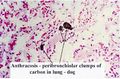 Anthracosis histo.jpg 395 × 261; 27 KB
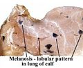 Melanosis gross.jpg 415 × 339; 27 KB
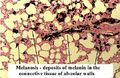 Melanosis histo.jpg 394 × 255; 34 KB
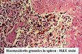 Haemosiderin.jpg 388 × 257; 35 KB
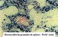 Haemosiderin prussian.jpg 391 × 255; 36 KB
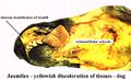 Jaundice discolouration.jpg 606 × 369; 38 KB
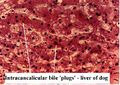 Jaundice bile plugs.jpg 374 × 266; 28 KB
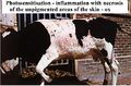 Photosensitisation.jpg 470 × 311; 37 KB
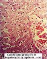
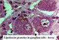 Lipofuscin granules.jpg 402 × 273; 28 KB
 Lipofuscin granules 2.jpg 314 × 397; 45 KB
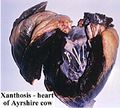 Lipofuscin xanthosis.jpg 417 × 375; 30 KB
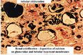 Renal calcification.jpg 369 × 242; 34 KB
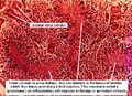 Urate crystals kidney.jpg 430 × 310; 50 KB
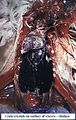 Urate crystals viscera.jpg 400 × 633; 59 KB
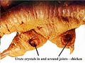 Urate crystals joints.jpg 429 × 319; 26 KB
 My horse.jpeg 480 × 640; 67 KB
 My dog and I.jpeg 640 × 480; 73 KB
 Cocktails.jpg 500 × 376; 30 KB
 Colon adhesions horse.jpg 801 × 535; 88 KB
 Pedunc lipoma closeup.jpg 816 × 519; 71 KB
 Large colon torsion horse.jpg 817 × 532; 110 KB
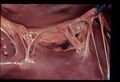 VSD1.jpg 1,157 × 790; 90 KB
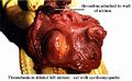 Thrombus cat left atrium.jpg 526 × 324; 31 KB
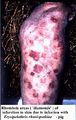 Skin infarction pig.jpg 395 × 624; 51 KB
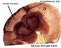 Renal infarction.jpg 546 × 437; 46 KB
 Thrombosis strongylus vulgaris.jpg 580 × 380; 49 KB
 VSD2.JPG 1,157 × 790; 110 KB
 VSD2.jpg 1,157 × 790; 110 KB
 Patent ductus goat.jpg 1,157 × 790; 99 KB
 Dextra-aorta.jpg 1,157 × 790; 111 KB
 Oligoden.png 675 × 600; 138 KB
 AV valve dysplasia cat.jpg 1,157 × 790; 91 KB
 Neuron.png 400 × 215; 32 KB
 Fibrinous pericarditis.jpg 1,157 × 790; 88 KB
 Traumatic pericarditis 2.jpg 1,157 × 790; 112 KB
 Ependyma1.jpg 400 × 267; 136 KB
 Choroid.jpg 400 × 267; 123 KB
 Traumatic pericarditis 4.jpg 1,157 × 790; 107 KB
 Traumatic reticulitis.jpg 1,157 × 790; 178 KB
 Pericarditis-histo.jpg 1,157 × 790; 160 KB
 Heart valve.jpg 1,157 × 790; 105 KB
 Endocardiosis2.jpg 1,157 × 790; 107 KB
 Endocardiosis3.jpg 1,157 × 790; 118 KB
 Endocardial calcification.jpg 1,157 × 790; 59 KB
 Bacterial endocarditis.jpg 1,157 × 790; 86 KB
 Parvovirus myocarditis.jpg 1,157 × 790; 147 KB
 Medial hypertrophy 2.jpg 1,157 × 790; 203 KB
 Arteriosclerosis.jpg 1,157 × 790; 115 KB
 Arterial hyalinisation.jpg 1,157 × 790; 167 KB
 Atheroma.jpg 1,157 × 790; 127 KB
 Endarteritis.jpg 1,157 × 790; 177 KB
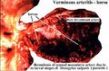
 Verminous arteritis.jpg 1,157 × 790; 83 KB
 Fibrinoid degeneration.jpg 1,157 × 790; 141 KB
 Dissecting aneurysm 2.jpg 1,157 × 790; 70 KB
 Dissecting aneurysm 3.jpg 1,157 × 790; 117 KB
 Pulmonary artery thrombus.jpg 1,157 × 790; 68 KB
 DIC thrombus.jpg 1,157 × 790; 183 KB
 Pulmonary oedema.jpg 477 × 349; 48 KB
 Pituitary dwarfism.jpg 1,157 × 790; 73 KB
 Pituitary cyst.jpg 1,157 × 790; 122 KB
 Pituitary tumour.jpg 1,157 × 790; 93 KB
 Normal thyroid.jpg 1,157 × 790; 97 KB
 Anasarca.jpg 335 × 227; 12 KB
 Parenchymatous goitre.jpg 1,157 × 790; 198 KB
 Colloid goitre.jpg 1,157 × 790; 123 KB
 Autoimmune thyroiditis.jpg 1,157 × 790; 184 KB
 Haemorrhage in the brain.jpg 291 × 389; 19 KB
 Thyroid carcinoma.jpg 1,157 × 790; 85 KB
 Bbb.gif 560 × 550; 51 KB
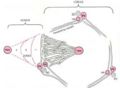
 Reflex-arc.gif 713 × 720; 40 KB
 Ascending-CNS.gif 647 × 530; 39 KB
 Neuron damage.jpg 2,311 × 3,318; 528 KB
 Neuron-damage.jpg 763 × 1,095; 92 KB
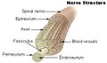 Nerve structure.jpg 338 × 200; 21 KB
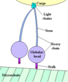 Cytoplasmic dynein.PNG 308 × 374; 8 KB
 Wallerian.jpg 870 × 740; 74 KB
 Myelination.gif 576 × 800; 34 KB
 001.JPG 324 × 219; 15 KB
 002.JPG 315 × 282; 29 KB
 03 - classic lobule.JPG 351 × 234; 19 KB
 04 - zones.JPG 315 × 232; 10 KB
 05 - hepatocyte.JPG 366 × 473; 42 KB
 06 - phagocytic cells (Kuppfer).JPG 204 × 318; 14 KB
 07 - Ito cells - lipocytes storing Vitamin A.JPG 264 × 174; 11 KB
 Normal adrenal cortex.jpg 1,157 × 790; 182 KB
 Adrenal atrophy.jpg 1,157 × 790; 160 KB
 Adrenal necrosis.jpg 1,157 × 790; 146 KB
 Adrenal necrosis2.jpg 1,157 × 790; 101 KB
 Cushings alopecia.jpg 1,157 × 790; 158 KB
 Chromophobe adenoma.jpg 1,157 × 790; 84 KB
 Nodular hyperplasia.jpg 1,157 × 790; 110 KB
 Adrenal neoplasia.jpg 1,157 × 790; 37 KB
 Mitotane therapy.jpg 1,157 × 790; 70 KB
 Myxoedema.jpg 1,157 × 790; 150 KB
 Flame follicles.jpg 1,157 × 790; 140 KB


.JPG)


