Unused files
Jump to navigation
Jump to search
The following files exist but are not embedded in any page. Please note that other web sites may link to a file with a direct URL, and so may still be listed here despite being in active use.
Showing below up to 100 results in range #301 to #400.
View (previous 100 | next 100) (20 | 50 | 100 | 250 | 500)
 Parvovirus dog.jpg 1,157 × 790; 131 KB
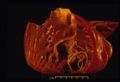
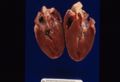 Hypertrophic cardiomyopathy.jpg 1,157 × 790; 57 KB
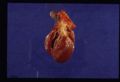
 Hypertrophic cardiomyopathy 2.jpg 1,157 × 790; 82 KB
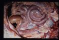
 Dirofilariasis.jpg 1,157 × 790; 69 KB
 Dirofilariasis 2.jpg 1,157 × 790; 72 KB
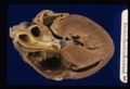
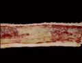
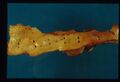 Aortic mineralisation.jpg 1,157 × 790; 65 KB
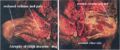
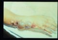
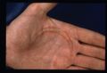 Hand burn injury.jpg 1,157 × 790; 90 KB
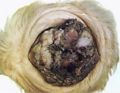
 Oral squamous cell carcinoma.jpg 1,157 × 790; 83 KB
 Thigh muscles atrophy.jpg 844 × 349; 71 KB
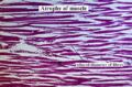 Muscle atrophy.jpg 374 × 247; 31 KB
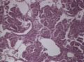
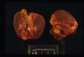 Thyroid adenoma.jpg 1,157 × 790; 76 KB
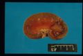
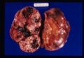 Renal carcinoma.jpg 1,157 × 790; 97 KB
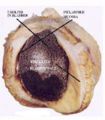
 Kidney infarct.jpg 1,157 × 790; 82 KB
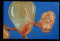 Hydronephrosis.jpg 1,157 × 790; 101 KB
 Human anthrax.jpg 1,157 × 790; 74 KB
 Obstructional hypertrophy bladder.jpg 364 × 415; 27 KB
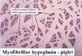 Myofibrillar hypoplasia.jpg 379 × 259; 25 KB
 Case pic 3.jpg 544 × 410; 79 KB
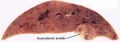 Hyperplastic nodule liver.jpg 768 × 271; 26 KB
 Benign prostatic hyperplasia.jpg 746 × 574; 89 KB
 Case pic 4.jpg 544 × 410; 78 KB
 Mammary tumour ossification.jpg 519 × 390; 59 KB
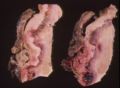
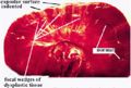 Renal dysplasia dog gross.jpg 423 × 284; 24 KB
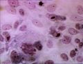
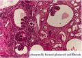 Renal dysplasia dog histological.jpg 386 × 274; 31 KB
 Anaplatic carcinoma.jpg 398 × 364; 41 KB
 Bronchiolitis.jpg 320 × 246; 18 KB
 COPD.jpg 320 × 247; 18 KB
 COPD scanning micrograph.jpg 320 × 246; 17 KB
 Dictyocaulus viviparus.jpg 320 × 245; 15 KB
 Diffuse fibrosing alveolitis.jpg 320 × 245; 13 KB
 Echinococcus cysts.jpg 320 × 244; 15 KB
 Fog fever 1.jpg 320 × 244; 11 KB
 Fog fever 2.jpg 320 × 245; 15 KB
 Interstitial emphysema.jpg 320 × 246; 13 KB
 Interstitial emphysema micro.jpg 320 × 240; 15 KB
 Pulmonary haemorrhage.jpg 320 × 242; 19 KB
 Pulmonary infarction.jpg 320 × 256; 20 KB
 Paraquat poisoning.jpg 320 × 246; 14 KB
 Parasitic bronchitis.jpg 320 × 239; 15 KB
 Parasitic pneumonia.jpg 320 × 233; 12 KB
 Pulmonary carcinoma.jpg 320 × 294; 19 KB
 Benign leiomyoma.jpg 222 × 631; 25 KB
 Perianal gland hepatoid adenoma dog.jpg 478 × 277; 37 KB
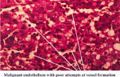
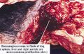 Haemangioscarcoma flank.jpg 556 × 360; 57 KB
 Haemangioscarcoma lung metastases.jpg 444 × 428; 38 KB
 Haemangioscarcoma histo.jpg 589 × 378; 58 KB
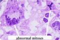 Abnormal mitoses.jpg 374 × 248; 17 KB
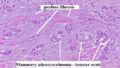 Profuse fibrosis.jpg 499 × 281; 37 KB
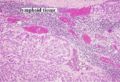 Adenocarcinoma metastasis to lymph node.jpg 432 × 294; 52 KB
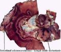 Intestinal adenocarcinoma lymphatic spread.jpg 520 × 443; 40 KB
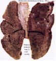 Melanoma metastases dog lung.jpg 426 × 464; 44 KB
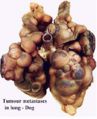 Tumour metastases lung dog.jpg 382 × 470; 30 KB
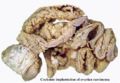 Coelomic implantation ovarian carcinoma.jpg 549 × 380; 36 KB
 Squamous cell carcinoma eye.jpg 394 × 304; 23 KB
 Squamous cell carcinoma histo.jpg 552 × 357; 61 KB
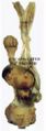
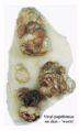 Viral papilloma.jpg 358 × 593; 27 KB
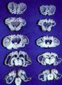 Swayback sections.jpg 361 × 494; 40 KB
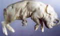 Cyclops.jpg 554 × 333; 35 KB
 Acute exudative pneumonia.jpg 320 × 246; 21 KB
 Acute exudative pneumonia - gross.jpg 320 × 242; 11 KB
 Acute fibrinous pneumonia.jpg 320 × 240; 14 KB
 Cleft palate.jpg 328 × 465; 21 KB
 Hydrocephalus.jpg 809 × 452; 67 KB
 Acute necrotising pneumonia.jpg 732 × 570; 55 KB
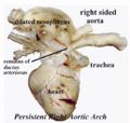 Persistent right aortic arch.jpg 471 × 447; 32 KB
 Acute suppurative pneumonia.jpg 742 × 574; 50 KB
 Adenomatosis of lung.jpg 744 × 544; 67 KB
 Adenovirus pneumonia.jpg 746 × 570; 50 KB
 Alveolar cell carcinoma.jpg 744 × 572; 83 KB
 Alveolar emphysema.jpg 758 × 574; 83 KB
 Aspergillus pneumonia of cattle.jpg 744 × 574; 63 KB
 Bronchiectasis.jpg 320 × 246; 20 KB
 Bronchiectasis micro.jpg 748 × 574; 103 KB
 Bronchiolitis obliterans.jpg 750 × 558; 63 KB
 Chronic bronchiolitis.jpg 746 × 572; 60 KB
 Thymus histo.jpg 758 × 540; 68 KB
 Peyers patches.jpg 758 × 556; 57 KB
 Chronic bronchopneumonia.jpg 750 × 564; 47 KB
 Calf pneumonia.jpg 746 × 554; 60 KB
 Collapsed trachea.jpg 742 × 576; 50 KB
 Contagious bovine pleuropneumonia.jpg 762 × 548; 48 KB
 Destructive emphysema - horse.jpg 752 × 574; 54 KB
 Enzootic pneumonia of pigs.jpg 742 × 570; 57 KB
 Fibrosarcoma in rhinarium of cat.jpg 744 × 576; 48 KB
 Gangrenous pneumonia.jpg 750 × 574; 59 KB
 Granulomatous mycotic pneumonia.jpg 746 × 576; 62 KB
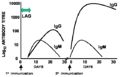 Primary and secondary response.jpg 561 × 362; 25 KB
 Guttural pouch mycosis.jpg 742 × 544; 54 KB
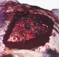
 Haemorrhage in GP mycosis.jpg 746 × 570; 68 KB
 Inclusion body rhinitis.jpg 744 × 576; 57 KB
 IBR nasal cavity.jpg 746 × 548; 70 KB
 IBR trachea.jpg 748 × 572; 38 KB
 Antibody structure.jpg 658 × 438; 34 KB
 Inhalation pneumonia.jpg 742 × 574; 89 KB
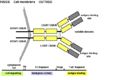
 TcR structure.jpg 399 × 279; 11 KB
 Interstitial pneumonia micro.jpg 748 × 480; 49 KB
 Laryngeal oedema.jpg 744 × 564; 49 KB
 Lipid pneumonia.jpg 744 × 570; 74 KB
 Lung carcinoma.jpg 742 × 552; 70 KB
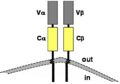
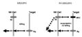 NK cell killing.jpg 604 × 270; 16 KB


