Unused files
The following files exist but are not embedded in any page. Please note that other web sites may link to a file with a direct URL, and so may still be listed here despite being in active use.
Showing below up to 100 results in range #251 to #350.
View (previous 100 | next 100) (20 | 50 | 100 | 250 | 500)
 Wallerian.jpg 870 × 740; 74 KB
 Myelination.gif 576 × 800; 34 KB
 001.JPG 324 × 219; 15 KB
 002.JPG 315 × 282; 29 KB
 03 - classic lobule.JPG 351 × 234; 19 KB
 04 - zones.JPG 315 × 232; 10 KB
 05 - hepatocyte.JPG 366 × 473; 42 KB
 06 - phagocytic cells (Kuppfer).JPG 204 × 318; 14 KB
 07 - Ito cells - lipocytes storing Vitamin A.JPG 264 × 174; 11 KB
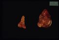
 Normal adrenal cortex.jpg 1,157 × 790; 182 KB
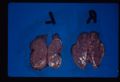
 Adrenal atrophy.jpg 1,157 × 790; 160 KB
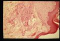
 Adrenal necrosis.jpg 1,157 × 790; 146 KB
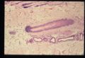
 Adrenal necrosis2.jpg 1,157 × 790; 101 KB
 Cushings alopecia.jpg 1,157 × 790; 158 KB
 Chromophobe adenoma.jpg 1,157 × 790; 84 KB
 Nodular hyperplasia.jpg 1,157 × 790; 110 KB
 Adrenal neoplasia.jpg 1,157 × 790; 37 KB
 Mitotane therapy.jpg 1,157 × 790; 70 KB
 Myxoedema.jpg 1,157 × 790; 150 KB
 Flame follicles.jpg 1,157 × 790; 140 KB
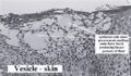 Serous fluid vesicle.jpg 527 × 306; 43 KB
 Acute fibrinous bronchopneumonia.jpg 639 × 328; 47 KB
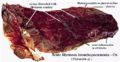
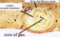 Acute abscess.jpg 393 × 242; 23 KB
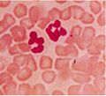 Neutrophils.jpg 310 × 281; 13 KB
 Eosinophils.jpg 322 × 270; 17 KB
 Mast cells.jpg 504 × 325; 37 KB
 Lymphocytes and plasma cell.jpg 391 × 258; 21 KB
 Macrophages.jpg 365 × 204; 15 KB
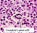 Langhans giant cell.jpg 531 × 485; 54 KB
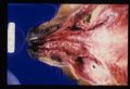
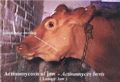 Lumpy jaw presentation.jpg 541 × 371; 41 KB
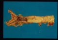
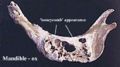 Lumpy jaw mandible.jpg 392 × 219; 17 KB
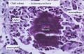 Lumpy jaw histology.jpg 403 × 259; 26 KB
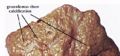 Tuberculous granulomas.jpg 462 × 215; 20 KB
 Perivascular cuffing.jpg 442 × 293; 44 KB
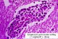
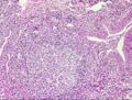 Lymphoid follicle lung.jpg 443 × 335; 64 KB
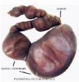 Pyometra.jpg 381 × 397; 23 KB
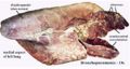 Bronchopneumonia ox.jpg 601 × 319; 40 KB
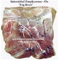 Fog fever.jpg 396 × 402; 33 KB
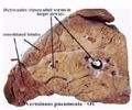 Lungworm.jpg 347 × 290; 24 KB
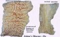 Johnes disease comparative.jpg 436 × 271; 23 KB
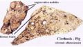 Cirrhosis pig.jpg 429 × 242; 25 KB
 Granultion tissue histology.jpg 742 × 574; 74 KB
 Fracture callus.jpg 742 × 574; 72 KB
 Vegetative endocarditis.jpg 1,157 × 790; 83 KB
 Fracture repair.jpg 744 × 534; 70 KB
 Scar tissue.jpg 746 × 556; 64 KB
 Dilated left atrium.jpg 1,157 × 790; 56 KB
 Thrombosis dog nose.jpg 1,157 × 790; 124 KB
 Thrombosis dog nose 2.jpg 1,157 × 790; 125 KB
 Sarcoma embolus.jpg 1,157 × 790; 74 KB
 Parvovirus dog.jpg 1,157 × 790; 131 KB
 Hypertrophic cardiomyopathy.jpg 1,157 × 790; 57 KB
 Hypertrophic cardiomyopathy 2.jpg 1,157 × 790; 82 KB
 Dirofilariasis.jpg 1,157 × 790; 69 KB
 Dirofilariasis 2.jpg 1,157 × 790; 72 KB
 Aortic mineralisation.jpg 1,157 × 790; 65 KB
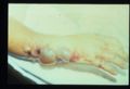
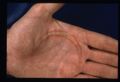 Hand burn injury.jpg 1,157 × 790; 90 KB
 Oral squamous cell carcinoma.jpg 1,157 × 790; 83 KB
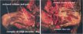 Thigh muscles atrophy.jpg 844 × 349; 71 KB
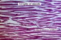 Muscle atrophy.jpg 374 × 247; 31 KB
 Thyroid adenoma.jpg 1,157 × 790; 76 KB
 Renal carcinoma.jpg 1,157 × 790; 97 KB
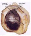
 Kidney infarct.jpg 1,157 × 790; 82 KB
 Hydronephrosis.jpg 1,157 × 790; 101 KB
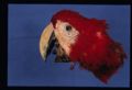
 Human anthrax.jpg 1,157 × 790; 74 KB
 Obstructional hypertrophy bladder.jpg 364 × 415; 27 KB
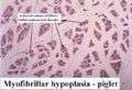 Myofibrillar hypoplasia.jpg 379 × 259; 25 KB
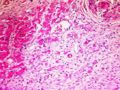 Case pic 3.jpg 544 × 410; 79 KB
 Hyperplastic nodule liver.jpg 768 × 271; 26 KB
 Benign prostatic hyperplasia.jpg 746 × 574; 89 KB
 Case pic 4.jpg 544 × 410; 78 KB
 Mammary tumour ossification.jpg 519 × 390; 59 KB
 Renal dysplasia dog gross.jpg 423 × 284; 24 KB
 Renal dysplasia dog histological.jpg 386 × 274; 31 KB
 Anaplatic carcinoma.jpg 398 × 364; 41 KB
 Bronchiolitis.jpg 320 × 246; 18 KB
 COPD.jpg 320 × 247; 18 KB
 COPD scanning micrograph.jpg 320 × 246; 17 KB
 Dictyocaulus viviparus.jpg 320 × 245; 15 KB
 Diffuse fibrosing alveolitis.jpg 320 × 245; 13 KB
 Echinococcus cysts.jpg 320 × 244; 15 KB
 Fog fever 1.jpg 320 × 244; 11 KB
 Fog fever 2.jpg 320 × 245; 15 KB
 Interstitial emphysema.jpg 320 × 246; 13 KB
 Interstitial emphysema micro.jpg 320 × 240; 15 KB
 Pulmonary haemorrhage.jpg 320 × 242; 19 KB
 Pulmonary infarction.jpg 320 × 256; 20 KB
 Paraquat poisoning.jpg 320 × 246; 14 KB
 Parasitic bronchitis.jpg 320 × 239; 15 KB
 Parasitic pneumonia.jpg 320 × 233; 12 KB
 Pulmonary carcinoma.jpg 320 × 294; 19 KB
 Benign leiomyoma.jpg 222 × 631; 25 KB
 Perianal gland hepatoid adenoma dog.jpg 478 × 277; 37 KB
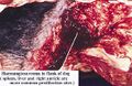 Haemangioscarcoma flank.jpg 556 × 360; 57 KB
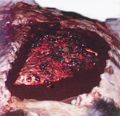 Haemangioscarcoma lung metastases.jpg 444 × 428; 38 KB
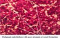 Haemangioscarcoma histo.jpg 589 × 378; 58 KB
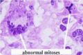 Abnormal mitoses.jpg 374 × 248; 17 KB
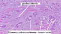 Profuse fibrosis.jpg 499 × 281; 37 KB
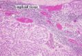 Adenocarcinoma metastasis to lymph node.jpg 432 × 294; 52 KB
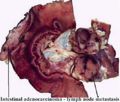 Intestinal adenocarcinoma lymphatic spread.jpg 520 × 443; 40 KB


.JPG)


