Uncategorized files
Showing below up to 250 results in range #101 to #350.
View (previous 250 | next 250) (20 | 50 | 100 | 250 | 500)
 Aspinall Slide1.JPG 960 × 720; 44 KB
 Aspinall Slide10.JPG 960 × 720; 71 KB
 Aspinall Slide11.JPG 960 × 720; 51 KB
 Aspinall Slide12.JPG 800 × 600; 51 KB
 Aspinall Slide13.JPG 960 × 720; 37 KB
 Aspinall Slide14.JPG 960 × 720; 60 KB
 Aspinall Slide15.JPG 960 × 720; 63 KB
 Aspinall Slide16.JPG 960 × 720; 38 KB
 Aspinall Slide2.JPG 960 × 720; 70 KB
 Aspinall Slide3.JPG 960 × 720; 76 KB
 Aspinall Slide4.JPG 960 × 720; 60 KB
 Aspinall Slide5.JPG 960 × 720; 72 KB
 Aspinall Slide6.JPG 960 × 720; 60 KB
 Aspinall Slide7.JPG 960 × 720; 82 KB
 Aspinall Slide8.JPG 960 × 720; 55 KB
 Aspinall Slide9.JPG 960 × 720; 57 KB
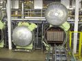 Atuoclav 2 (NXPowerLite Copy).JPG 4,000 × 3,000; 1.2 MB
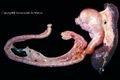
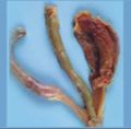
 Avian trachea with Syngamus trachea potcast.jpg 349 × 463; 29 KB
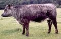
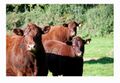
 Ayrshire.jpg 650 × 465; 75 KB
 BE5U9682 edit.jpg 5,184 × 3,456; 2.05 MB
 Bacteriology.png 436 × 156; 28 KB
- Error creating thumbnail: File missingBandera de españa.jpg 1,024 × 768; 68 KB
 Basic science.jpg 480 × 335; 56 KB
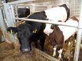 Belted galloway1.jpg 4,320 × 3,240; 3.05 MB
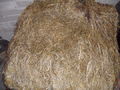 Big bale silage.jpg 4,320 × 3,240; 3.12 MB
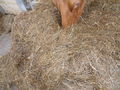 Big bale silage2.jpg 4,320 × 3,240; 3.09 MB
 Bioquimica.jpg 650 × 301; 28 KB
 Birds.png 436 × 156; 20 KB
 Bisected ovary from the mare potcast.jpg 630 × 311; 24 KB
 Bisecting angle positioning.jpg 667 × 482; 39 KB
 Bisecting angle technique.jpg 1,201 × 963; 78 KB
 Blood.png 436 × 156; 19 KB
 Blood Histology Dragster 1.jpg 600 × 450; 118 KB
 Blood Histology Dragster 2.jpg 600 × 450; 68 KB
 Blood Histology Dragster 3.jpg 600 × 450; 68 KB
 Blood Histology Dragster 4.jpg 600 × 450; 111 KB
 Blood profile.pdf ; 249 KB
 Bluefaced.jpg 640 × 473; 65 KB
 Bluefacedl.jpg 300 × 317; 23 KB
 Bluegrey.jpg 280 × 180; 18 KB
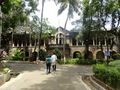 Bombay Veterinary College.jpg 180 × 135; 10 KB
 Bone and cartilage histology 1.jpg 600 × 450; 116 KB
 Bone and cartilate histology 2.jpg 600 × 450; 105 KB
 Bovine Pregnant Uterus.jpg 711 × 484; 91 KB
 Bovine Pregnant Uterus 2.jpg 666 × 482; 79 KB
 Bovine Viral Diarrhoea Virus (BVDV) - part 1.jpg 719 × 537; 68 KB
 Bovine Viral Diarrhoea Virus (BVDV) - part 2.jpg 714 × 517; 49 KB
 Bovine Viral Diarrhoea Virus (BVDV) - part 3.jpg 681 × 469; 92 KB
 Bovine liver potcast.jpg 720 × 351; 45 KB
 Bovine liver potcast 2.jpg 719 × 385; 65 KB
 Bovine omasum potcast.jpg 248 × 478; 28 KB
 Bovine reticular groove potcast.jpg 437 × 490; 68 KB
 Bovine teat and mammary gland potcast.jpg 719 × 496; 38 KB
 Bovine tongue with Taenia saginata potcast.jpg 667 × 491; 36 KB
 Brachyspira pilosicoli.jpg 401 × 304; 10 KB
 BristolCanineSkeleton.png 828 × 550; 176 KB
 Brown Manson cover.jpg 202 × 286; 11 KB
 Brushing cat teeth.jpg 662 × 562; 54 KB
 Brushing dog teeth.jpg 583 × 491; 26 KB
 Buddy graduate transparent.png 935 × 922; 88 KB
 BudrasCover.jpg 200 × 280; 9 KB
- Budras sample.pdf ; 608 KB
 Bugs.png 564 × 191; 23 KB
 ByVets.png 657 × 57; 5 KB
 CA.jpg 600 × 251; 221 KB
 CAT model part 1.jpg 1,688 × 858; 88 KB
 CAT model part 2.jpg 1,686 × 940; 141 KB
 CAT model part 3.jpg 1,689 × 895; 109 KB
 CAT model part 4.jpg 1,772 × 995; 178 KB
 CRC logo small.png 186 × 63; 6 KB
 CVconcepcion.jpg 730 × 310; 296 KB
 Calf housing screenshot.png 1,898 × 1,010; 243 KB
 Camelids.png 436 × 156; 23 KB
 Canine - female reproductive.jpg 600 × 485; 123 KB
 Canine Abdomen Still.png 605 × 535; 46 KB
 Canine Infectious Diseases- Self-Assessment Color Review.jpg 457 × 648; 39 KB
 Canine PTH.jpg 1,545 × 1,229; 539 KB
 Canine Pelvic Radiograph.jpg 600 × 450; 31 KB
 Canine Right lat pelvis.jpg 600 × 450; 26 KB
 Canine VD radiograph.jpg 600 × 450; 35 KB
 Canine abdomen 1.jpg 600 × 450; 55 KB
 Canine abdomen 2.jpg 600 × 450; 88 KB
 Canine abdomen dissection 1.jpg 600 × 396; 90 KB
 Canine abdomen dissection 2.jpg 600 × 360; 82 KB
 Canine abdomen dissection 3.jpg 600 × 377; 105 KB
 Canine abdomen dissection 4.jpg 600 × 386; 113 KB
 Canine abdomen potcast.jpg 534 × 492; 40 KB
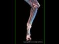
 Canine antebrachium.jpg 600 × 450; 43 KB
 Canine brain.jpg 600 × 450; 111 KB
 Canine carpus.JPG 600 × 450; 19 KB
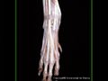
 Canine carpus and manus.jpg 600 × 450; 40 KB
 Canine carpus radiograph.jpg 600 × 450; 21 KB
 Canine cervical spine.jpg 600 × 450; 26 KB
 Canine cervical spine radiograph.jpg 600 × 450; 35 KB
 Canine cr-ca femur radiograph.jpg 600 × 450; 21 KB
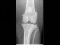 Canine cranio-caudal stifle radiograph.jpg 600 × 450; 16 KB
 Canine crus and pes 1.jpg 600 × 450; 51 KB
 Canine crus and pes 2.jpg 600 × 450; 36 KB
 Canine digestive system dissection.jpg 600 × 399; 68 KB
 Canine distal pelvic limb.jpg 600 × 450; 34 KB
 Canine dorsal skull 2.JPG 600 × 450; 17 KB
 Canine elbow.JPG 600 × 450; 15 KB
 Canine femur.jpg 600 × 450; 17 KB
 Canine flexed elbow radiograph.jpg 600 × 450; 28 KB
 Canine forelimb.jpg 600 × 450; 43 KB
 Canine forelimb craniolateral view.jpg 600 × 450; 40 KB
 Canine forelimb photo.jpg 600 × 450; 36 KB
 Canine frontal sinuses radiograph.jpg 600 × 450; 32 KB
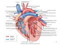
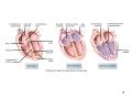
 Canine heart - left.jpg 600 × 486; 77 KB
 Canine heart - right.jpg 600 × 438; 70 KB
 Canine heart - sectioned.jpg 600 × 450; 88 KB
 Canine heart in situ.jpg 520 × 450; 118 KB
 Canine humerus.jpg 600 × 450; 25 KB
 Canine immature tarsus radiograph.jpg 600 × 450; 36 KB
 Canine intravenous ureterogram.jpg 600 × 450; 35 KB
 Canine larynx dissection.jpg 600 × 401; 96 KB
 Canine larynx radiograph.jpg 600 × 450; 45 KB
 Canine lateral abdomen radiograph.jpg 600 × 450; 44 KB
 Canine lateral femur radiograph.jpg 600 × 450; 20 KB
 Canine lateral pelvic radiograph.jpg 600 × 450; 42 KB
 Canine lateral skull 2.JPG 600 × 450; 25 KB
 Canine lateral skull radiograph.jpg 600 × 450; 43 KB
 Canine lateral stifle radiograph.jpg 600 × 450; 36 KB
 Canine lateral thoracic radiograph.jpg 600 × 450; 47 KB
 Canine lumbar spine 2.JPG 600 × 450; 30 KB
 Canine lumbar spine radiograph.jpg 600 × 450; 29 KB
 Canine male reproductive.jpg 600 × 430; 105 KB
 Canine male urethrogram radiograph.jpg 600 × 450; 49 KB
 Canine mandible.JPG 600 × 450; 20 KB
 Canine manus radiograph.jpg 600 × 450; 26 KB
 Canine metacarpus.JPG 600 × 450; 18 KB
 Canine metatarsus.JPG 600 × 450; 17 KB
 Canine nasal chamber2r.jpg 600 × 450; 49 KB
 Canine omega3 6.jpg 1,807 × 1,348; 662 KB
 Canine omega6.jpg 1,834 × 817; 458 KB
 Canine orbit and sagittal section of the canine head.jpg 715 × 494; 56 KB
 Canine palmar metacarpal.jpg 600 × 450; 18 KB
 Canine pelvic limb - deep dissection.jpg 477 × 450; 88 KB
 Canine pelvic limb - superficial dissection.jpg 421 × 450; 70 KB
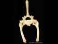
 Canine pelvis.jpg 600 × 450; 60 KB
 Canine pelvis 1.jpg 600 × 450; 55 KB
 Canine radius and ulna.JPG 600 × 450; 27 KB
 Canine sacrum.JPG 600 × 450; 24 KB
 Canine scapula.jpg 600 × 450; 24 KB
 Canine shoulder.JPG 600 × 450; 18 KB
 Canine shoulder.jpg 600 × 450; 64 KB
 Canine shoulder radiograph - cr-ca.jpg 600 × 450; 22 KB
 Canine shoulder radiograph - lateral.jpg 600 × 450; 28 KB
 Canine skull radiograph.jpg 600 × 450; 41 KB
 Canine stifle.JPG 600 × 450; 16 KB
 Canine stifle.jpg 465 × 450; 57 KB
 Canine tarsus.jpg 600 × 450; 17 KB
 Canine thoracic limb dissection 1.jpg 600 × 427; 103 KB
 Canine thoracic limb dissection 2.jpg 600 × 450; 50 KB
 Canine thoracic limb dissection 3.jpg 600 × 450; 78 KB
 Canine thoracic limb dissection 4.jpg 600 × 450; 44 KB
 Canine thoracic limb dissection 5.jpg 600 × 450; 39 KB
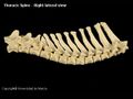 Canine thoracic spine.JPG 600 × 450; 26 KB
 Canine thoracic vert.jpg 600 × 450; 15 KB
 Canine thorax.jpg 600 × 450; 59 KB
 Canine thorax - deep.jpg 600 × 450; 55 KB
 Canine thorax - left 1.jpg 600 × 450; 40 KB
 Canine thorax - left 2.jpg 600 × 450; 45 KB
 Canine thorax - right 1.jpg 506 × 450; 103 KB
 Canine thorax - right 2.jpg 541 × 450; 120 KB
 Canine thorax - right 3.jpg 600 × 390; 120 KB
 Canine thorax and abdomen (left).jpg 600 × 450; 75 KB
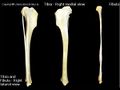 Canine tibia and fibula.JPG 600 × 450; 23 KB
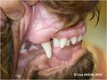 Canine tooth malocclusion.jpg 1,242 × 932; 88 KB
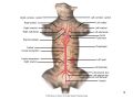
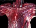 Canine ventral neck dissection.jpg 600 × 471; 120 KB
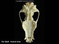 Canine ventral skull.jpg 600 × 450; 19 KB
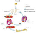 Canine vitamineD.jpg 1,445 × 1,340; 630 KB
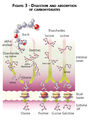 Carbohydrate digestibility.jpg 940 × 1,251; 513 KB
 Cardiovascular.png 564 × 191; 33 KB
 Cardiovascular 1 - heart wall.jpg 600 × 450; 77 KB
 Cardiovascular 2 - elastic artery.jpg 600 × 450; 124 KB
 Cardiovascular 3 - large vein.jpg 600 × 450; 66 KB
 Cardiovascular 4 - muscular artery.jpg 600 × 450; 69 KB
 Cardiovascular logo.png 340 × 340; 42 KB
- Care of the Orphan Donkey Foal.pdf ; 235 KB
 Case 12-image for Q11-image 4.jpg 512 × 512; 89 KB
 Case 12-image for Q3-image 2.JPG 350 × 400; 52 KB
 Case 12-image for Q7-image 3.JPG 512 × 512; 22 KB
 Case 12 North and Banks.jpg 386 × 400; 63 KB
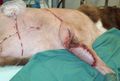
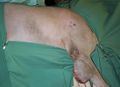
 Case 19-Q11-thoracodorsal draped in.JPG 988 × 781; 74 KB
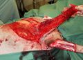 Case 19-Q11-thoracodorsal elevated flap.JPG 1,042 × 757; 133 KB
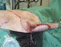
 Case 19-Q11-thoracodorsal end sx.JPG 1,132 × 766; 105 KB
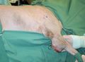
 Case 19-Q11-thoracodorsal end sx 2.JPG 1,105 × 853; 134 KB
 Case 19-Q11-thoracodorsal lines drawn.JPG 1,117 × 817; 94 KB
 Case 19-Q11-thoracodorsal prior to start.JPG 1,096 × 799; 97 KB
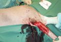 Case 19-Q11-thoracodorsal tumour out.JPG 1,054 × 739; 103 KB
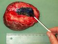 Case 19-Q9-specimen inked.JPG 1,280 × 960; 210 KB
 Case 19-Q9-specimen muscle surface.JPG 1,280 × 960; 213 KB
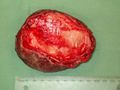
 Case 19-Q9-specimen skin surface.JPG 1,280 × 960; 162 KB
 Case 19-Q9-specimen sliced.JPG 1,280 × 960; 152 KB
 Case 22 IMG 0004 1.JPG 3,648 × 2,736; 4.85 MB
 Case 22 IMG 0006.JPG 3,648 × 2,736; 4.33 MB
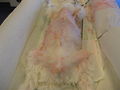 Case 22 IMG 0009.JPG 3,648 × 2,736; 3.69 MB
 Case 22 abdominal ultrasound.jpg 1,026 × 621; 169 KB
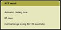 Case 22 act.jpg 644 × 319; 31 KB
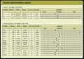 Case 22 biochemistry.jpg 987 × 694; 226 KB
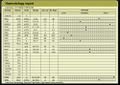 Case 22 haematology.jpg 997 × 706; 242 KB
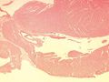
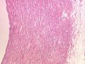
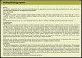 Case 22 histopathology.jpg 1,016 × 719; 333 KB
 Case 22 pcv and tp.jpg 768 × 181; 23 KB
 Case 22 urinalysis.jpg 771 × 233; 43 KB
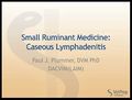 Caseous Lymphadenitis electure.jpg 617 × 468; 43 KB
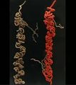 Cast of the testicular blood vessels potcast.jpg 427 × 480; 31 KB
- Castration donkey.pdf ; 133 KB
 Cat.png 436 × 156; 20 KB
- Cat Dental Decision Guide.pdf ; 479 KB
 Cat mandibular radiograph 1.jpg 631 × 494; 32 KB
 Cat mandibular radiograph 2.jpg 587 × 494; 31 KB
 Cat relationships example.jpg 1,959 × 1,450; 254 KB
 Cattle.png 436 × 156; 21 KB
 Ceva logo.jpg 144 × 140; 10 KB
 Chappie Dog Food Ad in the Daily Mail UK 5January1940.jpg 1,143 × 791; 481 KB
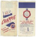 Chappie Dry Dog Food Bag 1935-36.jpg 815 × 821; 223 KB
 Charollais.gif 400 × 267; 79 KB
 Chicken Caeca Infected with Eimeria tenella.jpg 394 × 389; 27 KB
 Chinchilla-logo.png 300 × 305; 7 KB
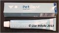 Chlorehexidine gel.jpg 541 × 301; 21 KB
 Chlorehexidine rinse.jpg 210 × 680; 16 KB
 Chris Palgrave.jpg 128 × 170; 29 KB
 ChungbukNU 01.jpg 600 × 445; 336 KB
 ChungbukNU 02.jpg 600 × 448; 243 KB
 Cimer.TIF 457 × 456; 289 KB
 Cimer.jpg 457 × 456; 109 KB
 Circulating hot air.jpg 960 × 642; 72 KB
 Cleft lip puppy.jpg 1,100 × 1,083; 78 KB
 Cleft palate 2.jpg 923 × 1,089; 93 KB
 Clinica-chillan.jpg 340 × 255; 46 KB
 Clinical-2.png 436 × 156; 21 KB
 Clinical.png 564 × 191; 34 KB
 Clinical skills - Applying a modified Robert Jones bandage.jpg 620 × 473; 58 KB
- Error creating thumbnail: File missingClinical skills - How to ‘closed glove’.jpg 665 × 471; 58 KB
- Error creating thumbnail: File missingClinical skills - How to ‘open glove’.jpg 662 × 471; 44 KB
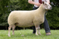
 ClunForest.jpg 709 × 531; 353 KB
 Clun forest.jpg 4,320 × 3,240; 3.14 MB
 CollvetUPLublin.jpg 2,000 × 3,008; 1.39 MB
 Colostrum hygiene screenshot.png 1,797 × 867; 1.56 MB
 Colostrum management screenshot.png 1,549 × 866; 328 KB
 Combined endodontic and periodontic lesion.jpg 1,183 × 844; 52 KB
 Comparative anatomy.png 640 × 428; 336 KB
 Comparison normal and necrotic Peyer's Patch.jpg 362 × 283; 22 KB
 Condylar process fracture.jpg 1,372 × 1,002; 133 KB
 Connective Tissue Dragster 1.jpg 598 × 450; 97 KB
 Connective Tissue Dragster 2.jpg 600 × 450; 99 KB
 Connective Tissue Dragster 3.jpg 600 × 450; 134 KB
 Contra-angle hand piece.jpg 484 × 892; 65 KB


.JPG)


_-_part_1.jpg)
_-_part_2.jpg)
_-_part_3.jpg)


.jpg)


