Uncategorized files
Showing below up to 100 results in range #201 to #300.
View (previous 100 | next 100) (20 | 50 | 100 | 250 | 500)
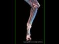
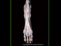 Canine distal pelvic limb.jpg 600 × 450; 34 KB
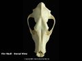 Canine dorsal skull 2.JPG 600 × 450; 17 KB
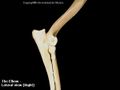 Canine elbow.JPG 600 × 450; 15 KB
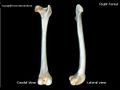 Canine femur.jpg 600 × 450; 17 KB
 Canine flexed elbow radiograph.jpg 600 × 450; 28 KB
 Canine forelimb.jpg 600 × 450; 43 KB
 Canine forelimb craniolateral view.jpg 600 × 450; 40 KB
 Canine forelimb photo.jpg 600 × 450; 36 KB
 Canine frontal sinuses radiograph.jpg 600 × 450; 32 KB
 Canine heart - left.jpg 600 × 486; 77 KB
 Canine heart - right.jpg 600 × 438; 70 KB
 Canine heart - sectioned.jpg 600 × 450; 88 KB
 Canine heart in situ.jpg 520 × 450; 118 KB
 Canine humerus.jpg 600 × 450; 25 KB
 Canine immature tarsus radiograph.jpg 600 × 450; 36 KB
 Canine intravenous ureterogram.jpg 600 × 450; 35 KB
 Canine larynx dissection.jpg 600 × 401; 96 KB
 Canine larynx radiograph.jpg 600 × 450; 45 KB
 Canine lateral abdomen radiograph.jpg 600 × 450; 44 KB
 Canine lateral femur radiograph.jpg 600 × 450; 20 KB
 Canine lateral pelvic radiograph.jpg 600 × 450; 42 KB
 Canine lateral skull 2.JPG 600 × 450; 25 KB
 Canine lateral skull radiograph.jpg 600 × 450; 43 KB
 Canine lateral stifle radiograph.jpg 600 × 450; 36 KB
 Canine lateral thoracic radiograph.jpg 600 × 450; 47 KB
 Canine lumbar spine 2.JPG 600 × 450; 30 KB
 Canine lumbar spine radiograph.jpg 600 × 450; 29 KB
 Canine male reproductive.jpg 600 × 430; 105 KB
 Canine male urethrogram radiograph.jpg 600 × 450; 49 KB
 Canine mandible.JPG 600 × 450; 20 KB
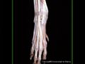
 Canine manus radiograph.jpg 600 × 450; 26 KB
 Canine metacarpus.JPG 600 × 450; 18 KB
 Canine metatarsus.JPG 600 × 450; 17 KB
 Canine nasal chamber2r.jpg 600 × 450; 49 KB
 Canine omega3 6.jpg 1,807 × 1,348; 662 KB
 Canine omega6.jpg 1,834 × 817; 458 KB
 Canine orbit and sagittal section of the canine head.jpg 715 × 494; 56 KB
 Canine palmar metacarpal.jpg 600 × 450; 18 KB
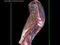
 Canine pelvic limb - deep dissection.jpg 477 × 450; 88 KB
 Canine pelvic limb - superficial dissection.jpg 421 × 450; 70 KB
 Canine pelvis.jpg 600 × 450; 60 KB
 Canine pelvis 1.jpg 600 × 450; 55 KB
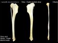
 Canine radius and ulna.JPG 600 × 450; 27 KB
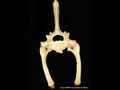
 Canine sacrum.JPG 600 × 450; 24 KB
 Canine scapula.jpg 600 × 450; 24 KB
 Canine shoulder.JPG 600 × 450; 18 KB
 Canine shoulder.jpg 600 × 450; 64 KB
 Canine shoulder radiograph - cr-ca.jpg 600 × 450; 22 KB
 Canine shoulder radiograph - lateral.jpg 600 × 450; 28 KB
 Canine skull radiograph.jpg 600 × 450; 41 KB
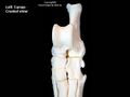
 Canine stifle.JPG 600 × 450; 16 KB
 Canine stifle.jpg 465 × 450; 57 KB
 Canine tarsus.jpg 600 × 450; 17 KB
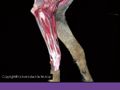
 Canine thoracic limb dissection 1.jpg 600 × 427; 103 KB
 Canine thoracic limb dissection 2.jpg 600 × 450; 50 KB
 Canine thoracic limb dissection 3.jpg 600 × 450; 78 KB
 Canine thoracic limb dissection 4.jpg 600 × 450; 44 KB
 Canine thoracic limb dissection 5.jpg 600 × 450; 39 KB
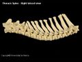 Canine thoracic spine.JPG 600 × 450; 26 KB
 Canine thoracic vert.jpg 600 × 450; 15 KB
 Canine thorax.jpg 600 × 450; 59 KB
 Canine thorax - deep.jpg 600 × 450; 55 KB
 Canine thorax - left 1.jpg 600 × 450; 40 KB
 Canine thorax - left 2.jpg 600 × 450; 45 KB
 Canine thorax - right 1.jpg 506 × 450; 103 KB
 Canine thorax - right 2.jpg 541 × 450; 120 KB
 Canine thorax - right 3.jpg 600 × 390; 120 KB
 Canine thorax and abdomen (left).jpg 600 × 450; 75 KB
 Canine tibia and fibula.JPG 600 × 450; 23 KB
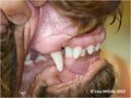 Canine tooth malocclusion.jpg 1,242 × 932; 88 KB
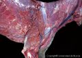
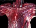 Canine ventral neck dissection.jpg 600 × 471; 120 KB
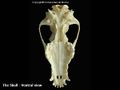 Canine ventral skull.jpg 600 × 450; 19 KB
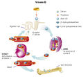 Canine vitamineD.jpg 1,445 × 1,340; 630 KB
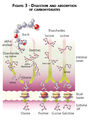 Carbohydrate digestibility.jpg 940 × 1,251; 513 KB
 Cardiovascular.png 564 × 191; 33 KB
 Cardiovascular 1 - heart wall.jpg 600 × 450; 77 KB
 Cardiovascular 2 - elastic artery.jpg 600 × 450; 124 KB
 Cardiovascular 3 - large vein.jpg 600 × 450; 66 KB
 Cardiovascular 4 - muscular artery.jpg 600 × 450; 69 KB
 Cardiovascular logo.png 340 × 340; 42 KB
 Care of the Orphan Donkey Foal.pdf ; 235 KB
 Case 12-image for Q11-image 4.jpg 512 × 512; 89 KB
 Case 12-image for Q3-image 2.JPG 350 × 400; 52 KB
 Case 12-image for Q7-image 3.JPG 512 × 512; 22 KB
 Case 12 North and Banks.jpg 386 × 400; 63 KB
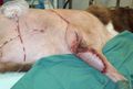
 Case 19-Q11-thoracodorsal draped in.JPG 988 × 781; 74 KB
 Case 19-Q11-thoracodorsal elevated flap.JPG 1,042 × 757; 133 KB
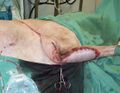
 Case 19-Q11-thoracodorsal end sx.JPG 1,132 × 766; 105 KB
 Case 19-Q11-thoracodorsal end sx 2.JPG 1,105 × 853; 134 KB
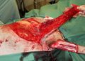
 Case 19-Q11-thoracodorsal lines drawn.JPG 1,117 × 817; 94 KB
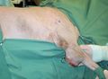
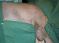 Case 19-Q11-thoracodorsal prior to start.JPG 1,096 × 799; 97 KB
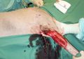 Case 19-Q11-thoracodorsal tumour out.JPG 1,054 × 739; 103 KB
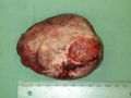
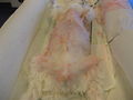
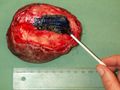 Case 19-Q9-specimen inked.JPG 1,280 × 960; 210 KB
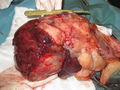
 Case 19-Q9-specimen muscle surface.JPG 1,280 × 960; 213 KB
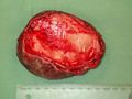
 Case 19-Q9-specimen skin surface.JPG 1,280 × 960; 162 KB
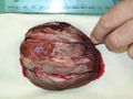 Case 19-Q9-specimen sliced.JPG 1,280 × 960; 152 KB
 Case 22 IMG 0004 1.JPG 3,648 × 2,736; 4.85 MB
 Case 22 IMG 0006.JPG 3,648 × 2,736; 4.33 MB
 Case 22 IMG 0009.JPG 3,648 × 2,736; 3.69 MB
 Case 22 abdominal ultrasound.jpg 1,026 × 621; 169 KB


.jpg)


