Search results
Jump to navigation
Jump to search
Page title matches
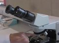
File:Using a microscope.jpg (665 × 480 (51 KB)) - 15:16, 27 September 2016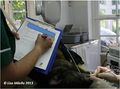
File:Filling a chart.jpg (945 × 699 (64 KB)) - 14:20, 6 August 2013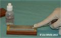
File:Sharpening a luxator.jpg (787 × 492 (31 KB)) - 14:21, 6 August 2013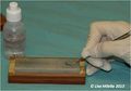
File:Sharpening a curette.jpg (775 × 541 (34 KB)) - 14:22, 6 August 2013
File:Saving a life.jpg (2,592 × 1,944 (1.2 MB)) - 15:59, 23 April 2012
File:Editing a WikiVet Page.pdf (859 KB) - 20:02, 25 January 2012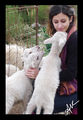
File:Anna Katogiritis A moment with lambs.jpg (1,282 × 1,860 (1.05 MB)) - 05:30, 8 June 2012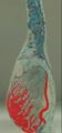
File:Isolated testis from a bull potcast.jpg (233 × 499 (21 KB)) - 14:21, 23 September 2016
File:How to perform a blood smear.jpg (650 × 435 (46 KB)) - 14:45, 27 September 2016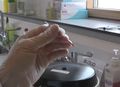
File:Obtain a Packed Cell Volume .jpg (664 × 482 (62 KB)) - 14:55, 27 September 2016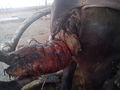
File:Postpartem prolapse in a She buffalo.jpg (2,560 × 1,920 (727 KB)) - 16:47, 17 January 2015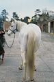
File:Equine Orthopaedics and Rheumatology Q&A 06.jpg (460 × 694 (164 KB)) - 13:01, 29 June 2012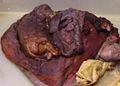
File:Female genital tract from a pregnant mare dissection.jpg (668 × 480 (71 KB)) - 13:57, 23 September 2016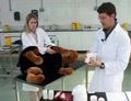
File:Clinical skills - Applying a modified Robert Jones bandage.jpg (620 × 473 (58 KB)) - 15:27, 26 September 2016File:Vibrio parahaemolyticus- An emerging foodborne pathogen-A Review.pdf (222 KB) - 10:58, 24 December 2011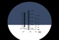
File:Obtain a Urine Specific Gravity (USG) and dipstick reading.jpg (650 × 445 (26 KB)) - 15:06, 27 September 2016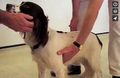
File:A guide to the cardiorespiratory examination of small animals.jpg (713 × 462 (62 KB)) - 14:34, 13 September 2016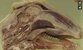
File:Lateral surface and sagittal section of the head of a sheep.jpg (710 × 434 (82 KB)) - 13:54, 13 September 2016


_and_dipstick_reading.jpg)


Page text matches

File:Dogandwikivet.jpg (448 × 745 (57 KB)) - 16:14, 30 March 2012
File:WikiPets3.jpg (466 × 392 (104 KB)) - 10:44, 15 June 2012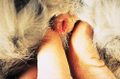
File:Manson SM17b.jpg (500 × 331 (139 KB)) - 13:20, 20 June 2011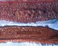
File:Comparison normal and necrotic Peyer's Patch.jpg (362 × 283 (22 KB)) - 16:40, 6 July 2011
File:Equine Abdominal Anatomy (left).jpg (600 × 450 (39 KB)) - 15:13, 15 June 2011
File:Equine Abdominal Anatomy (right).jpg (600 × 450 (56 KB)) - 09:40, 18 June 2011
File:Equine Forelimb Anatomy.jpg (600 × 450 (37 KB)) - 09:10, 18 June 2011
File:Equine Ventral Neck Anatomy.jpg (600 × 450 (62 KB)) - 09:13, 18 June 2011
File:Equine Forelimb Skeleton Anatomy.jpg (600 × 450 (14 KB)) - 09:14, 18 June 2011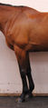
File:Equine Shoulder and Brachium Anatomy.jpg (268 × 600 (156 KB)) - 09:15, 18 June 2011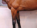
File:Equine Antebrachium Anatomy.jpg (600 × 450 (42 KB)) - 09:08, 18 June 2011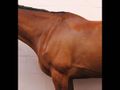
File:Equine Shoulder Anatomy.jpg (600 × 450 (22 KB)) - 09:15, 18 June 2011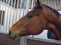
File:Equine head and neck.jpg (600 × 450 (55 KB)) - 18:02, 12 June 2011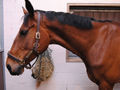
File:Equine head anatomy.jpg (600 × 450 (56 KB)) - 18:09, 12 June 2011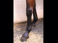
File:Equine Carpus and Manus Anatomy.jpg (600 × 450 (45 KB)) - 09:08, 18 June 2011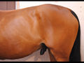
File:Equine Abdomen and Hip Anatomy.jpg (600 × 450 (45 KB)) - 11:22, 20 June 2011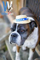
File:WikiPets10.jpg (481 × 720 (81 KB)) - 07:33, 20 June 2012
File:Equine Pelvic and Genital Anatomy.jpg (600 × 450 (35 KB)) - 12:16, 20 June 2011
File:Equine Pelvic Limb Anatomy.jpg (600 × 450 (19 KB)) - 11:26, 20 June 2011
File:Equine Pelvic and Femoral Anatomy.jpg (600 × 450 (44 KB)) - 12:12, 20 June 2011


.jpg)
.jpg)


