Search results
Jump to navigation
Jump to search
Page title matches
File:Using a microscope.jpg (665 × 480 (51 KB)) - 15:16, 27 September 2016
File:Saving a life.jpg (2,592 × 1,944 (1.2 MB)) - 15:59, 23 April 2012
File:Filling a chart.jpg |Description ={{en|1=Filling in a dental chart during examination}}(945 × 699 (64 KB)) - 14:20, 6 August 2013
File:Sharpening a luxator.jpg |Description ={{en|1=Sharpening a luxator on a flat stone}}(787 × 492 (31 KB)) - 14:21, 6 August 2013
File:Sharpening a curette.jpg |Description ={{en|1=Sharpening a curette on a flat stone }}(775 × 541 (34 KB)) - 14:22, 6 August 2013
File:Editing a WikiVet Page.pdf |Description ={{en|1=Basic instructions how to edit a WikiVet page.}}(859 KB) - 20:02, 25 January 2012
File:Isolated testis from a bull potcast.jpg (233 × 499 (21 KB)) - 14:21, 23 September 2016
File:Anna Katogiritis A moment with lambs.jpg ...iption={{en|1=This photo was taken on Karpathos island in Greece. While on a road trip, this lambs were on our way. They were left out of the fence whil(1,282 × 1,860 (1.05 MB)) - 05:30, 8 June 2012
File:How to perform a blood smear.jpg (650 × 435 (46 KB)) - 14:45, 27 September 2016
File:Obtain a Packed Cell Volume .jpg (664 × 482 (62 KB)) - 14:55, 27 September 2016
File:Postpartem prolapse in a She buffalo.jpg |description={{en|1=Irreduceable complicated postpartem prolapse of Uterus in a She buffalo}}(2,560 × 1,920 (727 KB)) - 16:47, 17 January 2015
File:Female genital tract from a pregnant mare dissection.jpg (668 × 480 (71 KB)) - 13:57, 23 September 2016
File:Equine Orthopaedics and Rheumatology Q&A 06.jpg (460 × 694 (164 KB)) - 13:01, 29 June 2012File:Vibrio parahaemolyticus- An emerging foodborne pathogen-A Review.pdf (222 KB) - 10:58, 24 December 2011
File:Clinical skills - Applying a modified Robert Jones bandage.jpg (620 × 473 (58 KB)) - 15:27, 26 September 2016
File:Obtain a Urine Specific Gravity (USG) and dipstick reading.jpg (650 × 445 (26 KB)) - 15:06, 27 September 2016
File:A guide to the cardiorespiratory examination of small animals.jpg (713 × 462 (62 KB)) - 14:34, 13 September 2016
File:Lateral surface and sagittal section of the head of a sheep.jpg (710 × 434 (82 KB)) - 13:54, 13 September 2016


_and_dipstick_reading.jpg)


Page text matches
File:Manson avian med 8.jpg |Description ={{en|1=This is a radiograph of a parrot with a lung abscess}}(462 × 692 (197 KB)) - 11:31, 29 June 2011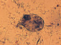
File:Manson SM 5.jpg |Description ={{en|1=This is a photo of a Sarcoptes mite from a mouse.}}(500 × 370 (267 KB)) - 09:55, 20 June 2011
File:Manson avian med 13.jpg |Description ={{en|1=This is a photo of a bird with a crop fistula}}(473 × 319 (128 KB)) - 12:57, 29 June 2011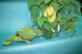
File:Manson avian med 7.jpg |Description ={{en|1=This is a photo of a parrot eating a house plant}}(481 × 318 (135 KB)) - 11:18, 29 June 2011
File:Manson SM 12.jpg |Description ={{en|1=This is a photo of a scent gland in a hamster.}}(500 × 295 (114 KB)) - 11:24, 20 June 2011
File:Manson avian med 14.jpg |Description ={{en|1=This is a photo of a fungally infected egg from a goose}}(475 × 317 (126 KB)) - 13:09, 29 June 2011
File:Manson SM 10.jpg |Description ={{en|1=This is a photo of a rabbit with a comminuted lumbar vertebral fracture.}}(500 × 310 (66 KB)) - 11:04, 20 June 2011
File:Manson avian med 18.jpg |Description ={{en|1=This is a photo of a cloacal urolith passed by a bird}}(500 × 333 (173 KB)) - 14:06, 29 June 2011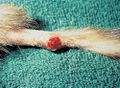
File:Manson SM 9.jpg |Description ={{en|1=This is a photo of a mast cell tumour on a ferrets tail}}(500 × 365 (203 KB)) - 10:50, 20 June 2011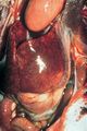
File:Manson avian med 11.jpg ...iption ={{en|1=This is a photo of a severely enlarged, mottled liver in a bird with hepatitis.}}(469 × 701 (312 KB)) - 12:25, 29 June 2011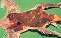
File:Manson avian med 19.jpg |Description ={{en|1=This is a photo showing a bird necropsy with a dark and congested liver and spleen}}(473 × 297 (156 KB)) - 14:17, 29 June 2011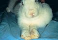
File:Manson SM15.jpg |Description ={{en|1=This is a photo of a rabbit with a nasal discharge and head tilt due to ''P. multocida'' infection.}}(500 × 340 (116 KB)) - 12:55, 20 June 2011
File:RedPin.png Pin used for Vetschools world map to denote a school without a page in WikiVet(15 × 26 (1 KB)) - 17:58, 24 November 2011
File:134957 Lat lumbar spine.jpg ...y of eating an unbalanced diet. The bone density is overall decreased with a poor corticomedullary distinction.}}(1,264 × 1,392 (389 KB)) - 14:33, 15 April 2015
File:SmAnOrth 03.jpg ...ph of the mouth of a cat which has undergone a stabilization procedure for a comminuted fracture of the angle of the left mandible.(500 × 337 (220 KB)) - 08:13, 2 September 2011
File:Tooth Extraction 5.jpg ...le flap used for tooth extraction. It is a mucogingival flap consisting of a sulcular incision and one vertical releasing incision. }}(1,302 × 869 (88 KB)) - 13:50, 1 May 2014
File:Equine Head Dissection (Sagittal).jpg |Description =Image of a dissection of a sagittal section of a horses head(600 × 450 (34 KB)) - 19:09, 22 June 2011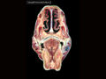
File:Equine Head Dissection (Transverse 1).jpg |Description =Image of a dissection of a transverse section of a horse´s head.(600 × 450 (37 KB)) - 19:22, 22 June 2011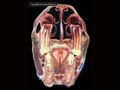
File:Equine Head Dissection (Transverse 2).jpg |Description =Image of a dissection of a transverse section of a horse´s head.(600 × 450 (186 KB)) - 19:19, 22 June 2011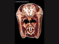
File:Equine Head Dissection (Transverse 3).jpg |Description =Image of a dissection of a transverse section of a horse´s head.(600 × 450 (43 KB)) - 19:20, 22 June 2011


.jpg)
.jpg)
.jpg)
.jpg)