Search results
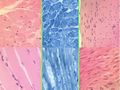
File:Muscle histology.jpg |Source =[[RVC|Royal Veterinary College]] |Author =[[RVC|Royal Veterinary College]](600 × 450 (139 KB)) - 18:34, 8 July 2011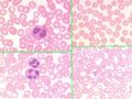
File:Blood Histology Dragster 1.jpg |Source =[[RVC|Royal Veterinary College]] |Author =[[RVC|Royal Veterinary College]](600 × 450 (118 KB)) - 16:33, 8 July 2011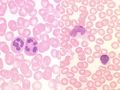
File:Blood Histology Dragster 2.jpg |Source =[[RVC|Royal Veterinary College]] |Author =[[RVC|Royal Veterinary College]](600 × 450 (68 KB)) - 17:04, 8 July 2011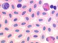
File:Blood Histology Dragster 3.jpg |Source =[[RVC|Royal Veterinary College]] |Author =[[RVC|Royal Veterinary College]](600 × 450 (68 KB)) - 17:06, 8 July 2011
File:Epithelia Dragster 1.jpg |Source =[[RVC|Royal Veterinary College]] |Author =[[RVC|Royal Veterinary College]](600 × 450 (115 KB)) - 19:28, 7 July 2011
File:Epithelia Dragster 2.jpg |Source =[[RVC|Royal Veterinary College]] |Author =[[RVC|Royal Veterinary College]](627 × 475 (113 KB)) - 19:30, 7 July 2011
File:Integument 2 - footpad.jpg |Source =[[RVC|Royal Veterinary College]] |Author =[[RVC|Royal Veterinary College]](600 × 450 (88 KB)) - 18:20, 8 July 2011
File:GIT 1 - oesophagus.jpg |Source =[[RVC|Royal Veterinary College]] |Author =[[RVC|Royal Veterinary College]](600 × 450 (92 KB)) - 17:45, 8 July 2011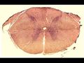
File:Spinal cord.jpg |Source =[[RVC|Royal Veterinary College]] |Author =[[RVC|Royal Veterinary College]](600 × 450 (52 KB)) - 19:46, 7 July 2011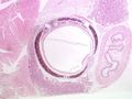
File:Respiratory 1 - trachea.jpg |Source =[[RVC|Royal Veterinary College]] |Author =[[RVC|Royal Veterinary College]](600 × 450 (82 KB)) - 17:53, 8 July 2011
File:Nerve cells 2.jpg |Source =[[RVC|Royal Veterinary College]] |Author =[[RVC|Royal Veterinary College]](600 × 450 (94 KB)) - 19:49, 7 July 2011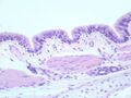
File:Respiratory 2 - lung.jpg |Source =[[RVC|Royal Veterinary College]] |Author =[[RVC|Royal Veterinary College]](600 × 450 (57 KB)) - 17:57, 8 July 2011
File:Cardiovascular 2 - elastic artery.jpg |Source =[[RVC|Royal Veterinary College]] |Author =[[RVC|Royal Veterinary College]](600 × 450 (124 KB)) - 18:05, 8 July 2011
File:Canine antebrachium.jpg |Source =[[RVC|Royal Veterinary College]] |Author =[[RVC|Royal Veterinary College]](600 × 450 (43 KB)) - 18:04, 6 July 2011
File:Cardiovascular 4 - muscular artery.jpg |Source =[[RVC|Royal Veterinary College]] |Author =[[RVC|Royal Veterinary College]](600 × 450 (69 KB)) - 18:09, 8 July 2011
File:Dog skeleton.jpg |Source =[[RVC|Royal Veterinary College]] |Author =[[RVC|Royal Veterinary College]](600 × 450 (19 KB)) - 11:01, 23 June 2011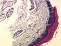
File:Integument 1 - digital skin.jpg |Source =[[RVC|Royal Veterinary College]] |Author =[[RVC|Royal Veterinary College]](600 × 450 (96 KB)) - 18:19, 8 July 2011
File:Connective Tissue Dragster 1.jpg |Source =[[RVC|Royal Veterinary College]] |Author =[[RVC|Royal Veterinary College]](598 × 450 (97 KB)) - 19:37, 7 July 2011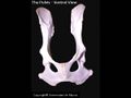
File:Ventral pelvis 2.jpg |Source =[[RVC|Royal Veterinary College]] |Author =[[RVC|Royal Veterinary College]](600 × 450 (18 KB)) - 12:26, 23 June 2011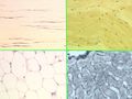
File:Connective Tissue Dragster 2.jpg |Source =[[RVC|Royal Veterinary College]] |Author =[[RVC|Royal Veterinary College]](600 × 450 (99 KB)) - 19:38, 7 July 2011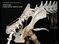
File:Dorsal pelvis 2.jpg |Source =[[RVC|Royal Veterinary College]] |Author =[[RVC|Royal Veterinary College]](600 × 450 (37 KB)) - 12:27, 23 June 2011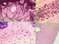
File:Connective Tissue Dragster 3.jpg |Source =[[RVC|Royal Veterinary College]] |Author =[[RVC|Royal Veterinary College]](600 × 450 (134 KB)) - 19:42, 7 July 2011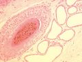
File:Integument 6 - skin with hair follicle.jpg |Source =[[RVC|Royal Veterinary College]] |Author =[[RVC|Royal Veterinary College]](600 × 450 (68 KB)) - 18:29, 8 July 2011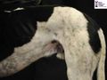
File:Canine pelvis 1.jpg |Source =[[RVC|Royal Veterinary College]] |Author =[[RVC|Royal Veterinary College]](600 × 450 (55 KB)) - 17:59, 6 July 2011
File:Cardiovascular 3 - large vein.jpg |Source =[[RVC|Royal Veterinary College]] |Author =[[RVC|Royal Veterinary College]](600 × 450 (66 KB)) - 18:06, 8 July 2011
File:Blood Histology Dragster 4.jpg |Source =[[RVC|Royal Veterinary College]] |Author =[[RVC|Royal Veterinary College]](600 × 450 (111 KB)) - 17:10, 8 July 2011
File:Bone and cartilage histology 1.jpg |Source =[[RVC|Royal Veterinary College]] |Author =[[RVC|Royal Veterinary College]](600 × 450 (116 KB)) - 17:18, 8 July 2011
File:Whole hindlimb 2.jpg |Source =[[RVC|Royal Veterinary College]] |Author =[[RVC|Royal Veterinary College]](600 × 450 (16 KB)) - 12:17, 23 June 2011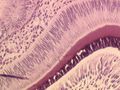
File:Oral cav 1.jpg |Source =[[RVC|Royal Veterinary College]] |Author =[[RVC|Royal Veterinary College]](600 × 450 (98 KB)) - 17:29, 8 July 2011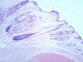
File:Integument 3 - hair follicle.jpg |Source =[[RVC|Royal Veterinary College]] |Author =[[RVC|Royal Veterinary College]](600 × 450 (74 KB)) - 18:22, 8 July 2011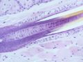
File:Integument 4 - hair follicle long section.jpg |Source =[[RVC|Royal Veterinary College]] |Author =[[RVC|Royal Veterinary College]](600 × 450 (66 KB)) - 18:24, 8 July 2011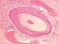
File:Integument 5 - sinus hair.jpg |Source =[[RVC|Royal Veterinary College]] |Author =[[RVC|Royal Veterinary College]](600 × 450 (88 KB)) - 18:25, 8 July 2011
File:Canine carpus and manus.jpg |Source = [[RVC|Royal Veterinary College]] |Author = [[RVC|Royal Veterinary College]](600 × 450 (40 KB)) - 18:05, 6 July 2011
File:Equine Thoracic and Abdominal Viscera Anatomy (right).jpg |Source =[[RVC|Royal Veterinary College]] |Author =[[RVC|Royal Veterinary College]](600 × 450 (57 KB)) - 09:20, 18 June 2011
File:Equine Thoracic and Abdominal Viscera Anatomy (left).jpg |Source =[[RVC|Royal Veterinary College]] |Author =[[RVC|Royal Veterinary College]](600 × 450 (44 KB)) - 09:17, 18 June 2011
File:Bone and cartilate histology 2.jpg |Source =[[RVC|Royal Veterinary College]] |Author =[[RVC|Royal Veterinary College]](600 × 450 (105 KB)) - 17:22, 8 July 2011
File:Oral Cavity Gland.jpg |Source =[[RVC|Royal Veterinary College]] |Author =[[RVC|Royal Veterinary College]](600 × 450 (163 KB)) - 17:38, 8 July 2011
File:Canine thorax and abdomen (left).jpg |Source =[[RVC|Royal Veterinary College]] |Author =[[RVC|Royal Veterinary College]](600 × 450 (75 KB)) - 17:49, 6 July 2011
File:Cardiovascular 1 - heart wall.jpg |Source =[[RVC|Royal Veterinary College]] |Author =[[RVC|Royal Veterinary College]](600 × 450 (77 KB)) - 18:03, 8 July 2011
File:Equine Abdominal Anatomy (right).jpg |Source =[[RVC|Royal Veterinary College]] |Author =[[RVC|Royal Veterinary College]](600 × 450 (56 KB)) - 09:40, 18 June 2011
File:Equine Antebrachium Anatomy.jpg |Source =[[RVC|Royal Veterinary College]] |Author =[[RVC|Royal Veterinary College]](600 × 450 (42 KB)) - 09:08, 18 June 2011
File:Equine Brain Anatomy (Transverse).jpg |Source =[[RVC|Royal Veterinary College]] |Author =[[RVC|Royal Veterinary College]](500 × 375 (69 KB)) - 07:27, 23 June 2011
File:Equine Forelimb Anatomy.jpg |Source =[[RVC|Royal Veterinary College]] |Author =[[RVC|Royal Veterinary College]](600 × 450 (37 KB)) - 09:10, 18 June 2011
File:Equine Ventral Neck Anatomy.jpg |Source =[[RVC|Royal Veterinary College]] |Author =[[RVC|Royal Veterinary College]](600 × 450 (62 KB)) - 09:13, 18 June 2011
File:Equine Forelimb Skeleton Anatomy.jpg |Source =[[RVC|Royal Veterinary College]] |Author =[[RVC|Royal Veterinary College]](600 × 450 (14 KB)) - 09:14, 18 June 2011
File:Equine Pelvic Limb Dissection Anatomy.jpg |Source =[[RVC|Royal Veterinary College]] |Author =[[RVC|Royal Veterinary College]](668 × 450 (34 KB)) - 10:54, 23 June 2011
File:Equine Shoulder and Brachium Anatomy.jpg |Source =[[RVC|Royal Veterinary College]] |Author =[[RVC|Royal Veterinary College]](268 × 600 (156 KB)) - 09:15, 18 June 2011
File:Equine Shoulder Anatomy.jpg |Source =[[RVC|Royal Veterinary College]] |Author =[[RVC|Royal Veterinary College]](600 × 450 (22 KB)) - 09:15, 18 June 2011
File:Equine Carpus and Manus Anatomy.jpg |Source =[[RVC|Royal Veterinary College]] |Author =[[RVC|Royal Veterinary College]](600 × 450 (45 KB)) - 09:08, 18 June 2011
File:Equine head and neck.jpg |Source =[[RVC|Royal Veterinary College]] |Author =[[RVC|Royal Veterinary College]](600 × 450 (55 KB)) - 18:02, 12 June 2011
File:Equine head anatomy.jpg |Source =[[RVC|Royal Veterinary College]] |Author =[[RVC|Royal Veterinary College]](600 × 450 (56 KB)) - 18:09, 12 June 2011
File:Equine Abdomen and Hip Anatomy.jpg |Source =[[RVC|Royal Veterinary College]] |Author =[[RVC|Royal Veterinary College]](600 × 450 (45 KB)) - 11:22, 20 June 2011
File:Whole dog - right.jpg |Source = [[RVC|Royal Veterinary College]] |Author = [[RVC|Royal Veterinary College]](600 × 450 (52 KB)) - 17:48, 6 July 2011
File:Equine Brain Anatomy (Ventral).jpg |Source =[[RVC|Royal Veterinary College]] |Author =[[RVC|Royal Veterinary College]](600 × 450 (48 KB)) - 07:26, 23 June 2011
File:Equine Thoracic Anatomy (right).jpg |Source =[[RVC|Royal Veterinary College]] |Author =[[RVC|Royal Veterinary College]](643 × 450 (79 KB)) - 10:43, 23 June 2011
File:Equine Head Dissection (Coronal).jpg |Source =[[RVC|Royal Veterinary College]] |Author =[[RVC|Royal Veterinary College]](600 × 450 (37 KB)) - 19:10, 22 June 2011
File:Equine Thoracic Anatomy (right w-out lung).jpg |Source =[[RVC|Royal Veterinary College]] |Author =[[RVC|Royal Veterinary College]](684 × 450 (237 KB)) - 10:43, 23 June 2011
File:Equine Head Dissection (lateral).gif |Source =[[RVC|Royal Veterinary College]] |Author =[[RVC|Royal Veterinary College]](600 × 491 (238 KB)) - 19:08, 22 June 2011
File:Equine Pelvic and Genital Anatomy.jpg |Source =[[RVC|Royal Veterinary College]] |Author =[[RVC|Royal Veterinary College]](600 × 450 (35 KB)) - 12:16, 20 June 2011
File:Equine Pelvic Limb Anatomy.jpg |Source =[[RVC|Royal Veterinary College]] |Author =[[RVC|Royal Veterinary College]](600 × 450 (19 KB)) - 11:26, 20 June 2011
File:Equine Abdominal Anatomy (left).jpg |Source =[[RVC|Royal Veterinary College]] |Author =[[RVC|Royal Veterinary College]](600 × 450 (39 KB)) - 15:13, 15 June 2011
File:Equine Pelvic and Femoral Anatomy.jpg |Source =[[RVC|Royal Veterinary College]] |Author =[[RVC|Royal Veterinary College]](600 × 450 (44 KB)) - 12:12, 20 June 2011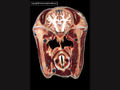
File:Equine Head Dissection (Transverse 3).jpg |Source =[[RVC|Royal Veterinary College]] |Author =[[RVC|Royal Veterinary College]](600 × 450 (43 KB)) - 19:20, 22 June 2011
File:Equine Head Dissection (Sagittal).jpg |Source =[[RVC|Royal Veterinary College]] |Author =[[RVC|Royal Veterinary College]](600 × 450 (34 KB)) - 19:09, 22 June 2011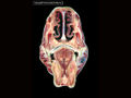
File:Equine Head Dissection (Transverse 1).jpg |Source =[[RVC|Royal Veterinary College]] |Author =[[RVC|Royal Veterinary College]](600 × 450 (37 KB)) - 19:22, 22 June 2011
File:Equine Head Dissection (Transverse 2).jpg |Source =[[RVC|Royal Veterinary College]] |Author =[[RVC|Royal Veterinary College]](600 × 450 (186 KB)) - 19:19, 22 June 2011
File:Equine Crus and Pes Anatomy (Cranial view).jpg |Source =[[RVC|Royal Veterinary College]] |Author =[[RVC|Royal Veterinary College]](600 × 450 (33 KB)) - 12:08, 20 June 2011
File:Equine Crus and Pes Anatomy.jpg |Source =[[RVC|Royal Veterinary College]] |Author =[[RVC|Royal Veterinary College]](600 × 450 (31 KB)) - 12:06, 20 June 2011
File:Heterophil.jpg |Source =RVC(240 × 180 (5 KB)) - 23:28, 21 August 2011
File:Museum1.jpeg |Description ={{en|1=RVC Museum}}(802 × 614 (57 KB)) - 07:49, 16 August 2011
File:Equine locomotion snapshot.png |Source=RVC(1,280 × 560 (371 KB)) - 13:11, 23 August 2011
File:Mix small.jpg |Source =RVC eMedia(150 × 100 (10 KB)) - 13:36, 19 September 2011
File:Diabetes mellitus e lecture.jpg |Source =RVC(1,040 × 774 (80 KB)) - 10:16, 13 November 2014
File:Mix-elearning.jpg |Source =RVC e-Media(400 × 267 (38 KB)) - 13:41, 19 September 2011
File:OVAM student workshop.JPG |Description ={{en|1=OVAM e-curator workshop delegates at the RVC}}(3,648 × 2,736 (2.76 MB)) - 21:41, 16 April 2012
File:Stubbs dragster.jpg |Permission =RVC(223 × 165 (12 KB)) - 19:10, 17 August 2011
File:Taurine-deficient cat.jpg |Source =RVC(509 × 412 (129 KB)) - 14:16, 15 April 2015
File:Kitty barty LVtxt.jpg |Source =RVC(501 × 408 (112 KB)) - 14:25, 15 April 2015
File:Mcleasby.jpg |description={{en|1=Mark Cleasby RVC}}(135 × 180 (30 KB)) - 14:56, 4 September 2014
File:174223 Right4.jpg |Source =RVC(1,280 × 1,024 (256 KB)) - 14:07, 15 April 2015
File:134957 Lat lumbar spine.jpg |Source =RVC(1,264 × 1,392 (389 KB)) - 14:33, 15 April 2015
File:Anatomía Radiográfica del Tórax.swf |source=[[RVC|Royal Veterinary College]](6.29 MB) - 07:38, 7 May 2012
File:Dragster logo.png |Source=RVC(213 × 188 (26 KB)) - 11:10, 1 July 2011File:Anatomía Radiográfica de la Columna Vertebral.swf |source=[[RVC|Royal Veterinary College]](3.58 MB) - 07:41, 7 May 2012File:ECase 1.swf |description={{en|1=An eCase from the RVC. An 18mo FE Lab X presents to a referral institute with a heart murmur dete(2.2 MB) - 11:08, 1 August 2012


.jpg)
.jpg)


.jpg)

.jpg)


.jpg)
.jpg)
.jpg)
.jpg)
.gif)


.jpg)

.jpg)
.jpg)


.jpg)
.jpg)
.jpg)
.jpg)