Uploads by Lizzies
This special page shows all uploaded files.
| Date | Name | Thumbnail | Size | Description | Versions |
|---|---|---|---|---|---|
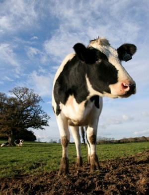
| 16:28, 7 August 2007 | Cow2.jpg (file) |  |
24 KB | 1 | |
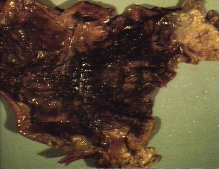
| 16:21, 9 August 2007 | Haemorrhagic gastritis.jpg (file) |  |
63 KB | 1 | |
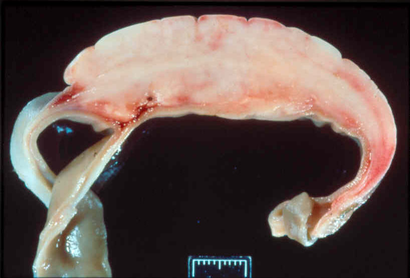
| 18:24, 9 August 2007 | Abomasal lymphoma.jpg (file) |  |
68 KB | 1 | |
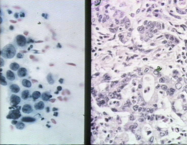
| 18:39, 9 August 2007 | Adenocarcinoma stomach histopath2.jpg (file) |  |
64 KB | 1 | |
| 18:39, 9 August 2007 | Adenocarcinoma stomach.jpg (file) |  |
69 KB | 1 | |
| 18:46, 9 August 2007 | Oesophageal bloat line.jpg (file) |  |
58 KB | 1 | |
| 18:52, 9 August 2007 | Traumatic pericarditis.jpg (file) |  |
7 KB | 1 | |
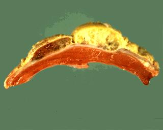
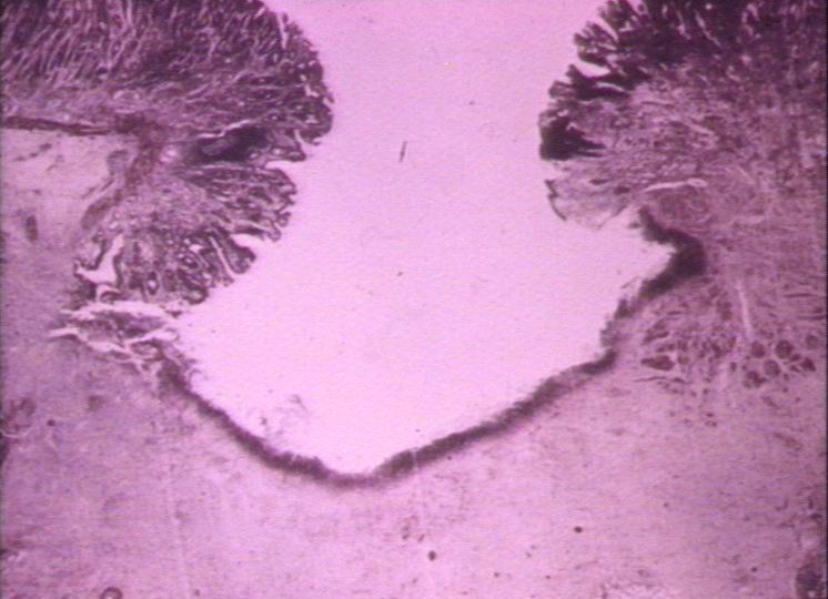
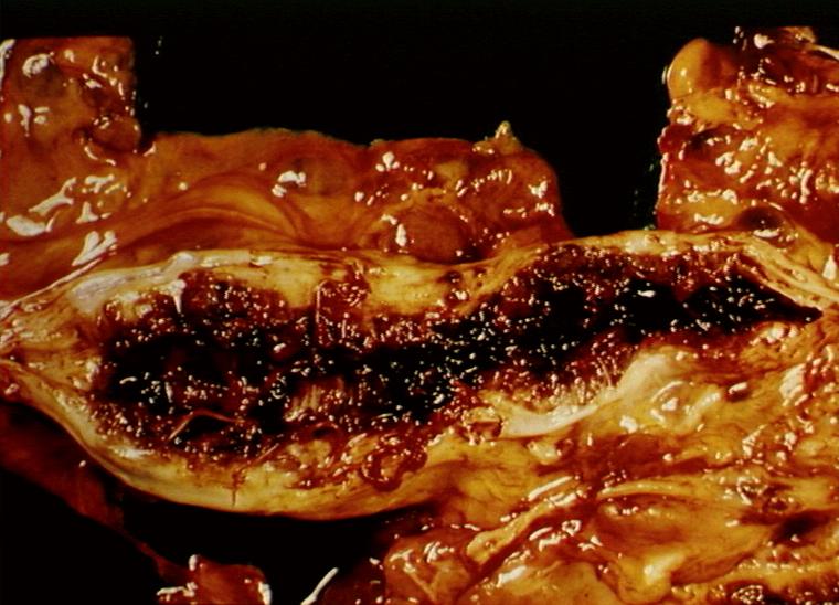
| 19:03, 9 August 2007 | Gastric ulcer.jpg (file) |  |
65 KB | Gross appearance of a gastric ulcer. | 1 |
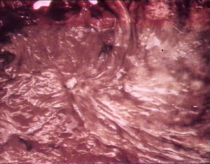
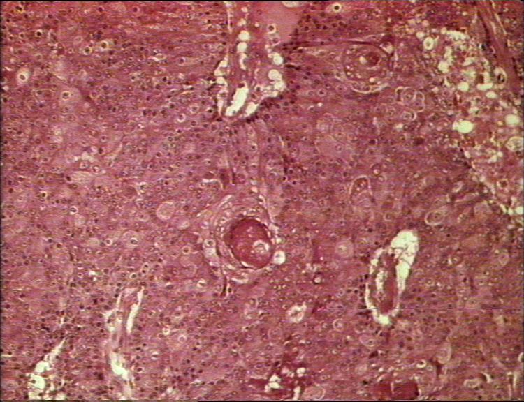
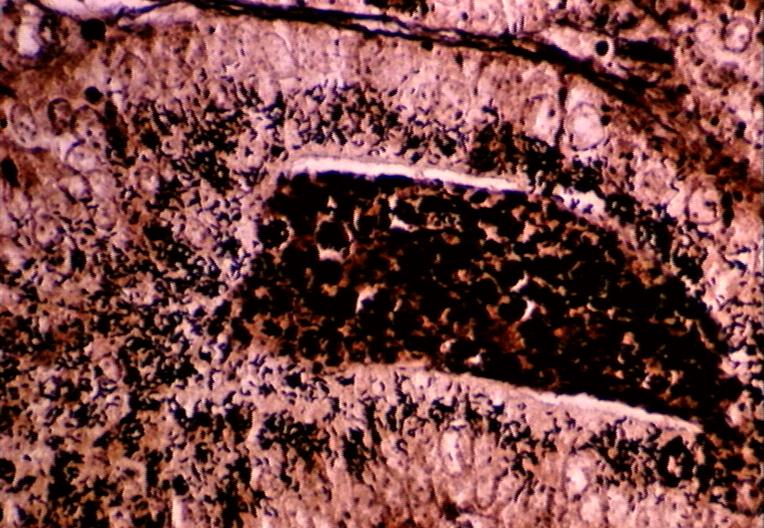
| 19:04, 9 August 2007 | Gastric ulcer histopath.jpg (file) |  |
55 KB | Histological section through a gastric ulcer. | 1 |
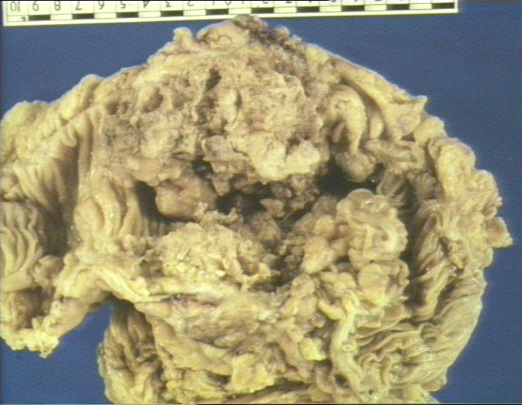
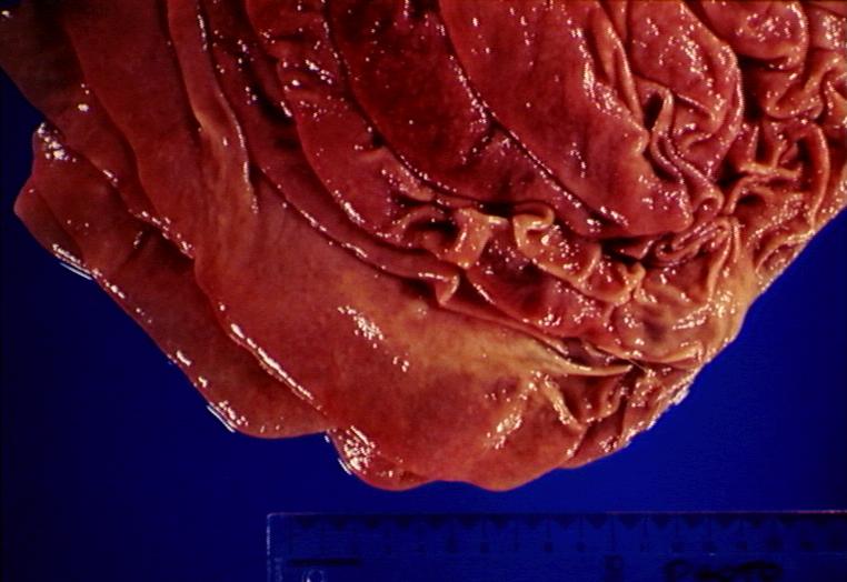
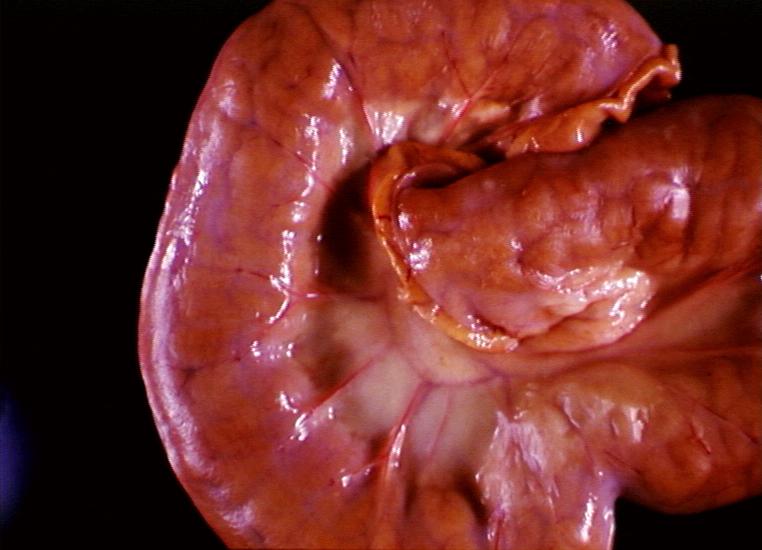
| 19:07, 9 August 2007 | Ostertagiasis.jpg (file) |  |
57 KB | Ostertagiosis caused by Ostertagia ostertagi: abomasum showing oedema and inflamation of fundic folds. | 1 |
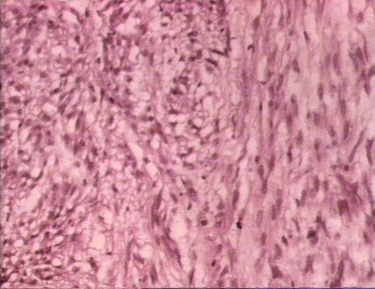
| 19:16, 9 August 2007 | Leiomyoma.jpg (file) |  |
67 KB | Histological appearance of leiomyoma. | 1 |
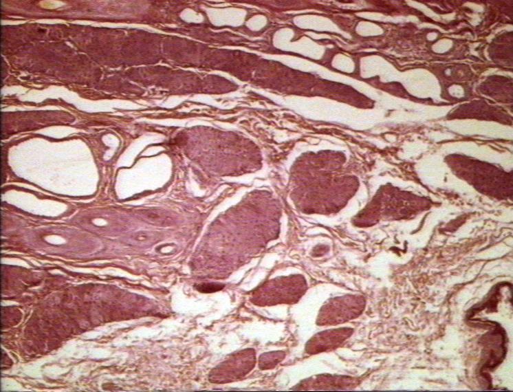
| 10:51, 10 August 2007 | Normal perianal gland.jpg (file) |  |
97 KB | Histological appearance of a normal perianal gland. | 1 |
| 10:51, 10 August 2007 | Perianal gland adenoma histopath.jpg (file) |  |
100 KB | Histological appearance of a perianal gland adenoma. | 1 |
| 10:52, 10 August 2007 | Perianal gland adenoma.jpg (file) |  |
51 KB | Gross appearance of a perianal gland adenoma. | 1 |
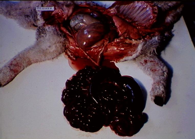
| 11:00, 10 August 2007 | Strongylus vulgaris.jpg (file) |  |
78 KB | Thrombosis of cranial mesenteric artery caused by Strongylus vulgaris larvae. | 1 |
| 11:15, 10 August 2007 | Infaction of the small bowel.jpg (file) |  |
65 KB | Infaction of the small bowel. | 1 |
| 11:24, 10 August 2007 | Brunner gland adenoma.jpg (file) |  |
75 KB | Adenoma of brunners gland (duodenum). | 1 |
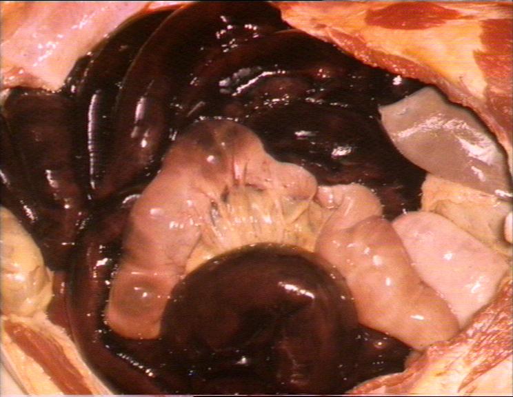
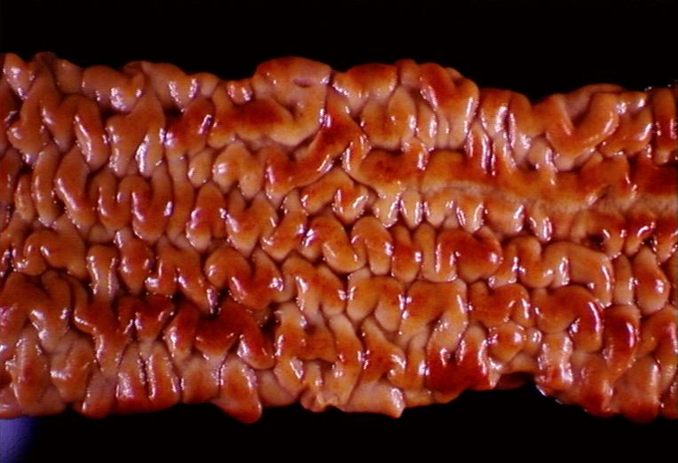
| 21:06, 13 August 2007 | Johnes disease proliferative enteritis.jpg (file) |  |
50 KB | Johnes disease - proliferative enteritis. | 1 |
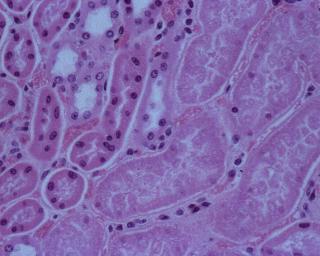
| 21:16, 13 August 2007 | Pulpy kidney disease.jpg (file) |  |
15 KB | Abrupt transition between viable and necrotic renal tubules in pulpy kidney disease. | 1 |
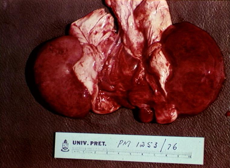
| 21:19, 13 August 2007 | Pulpy kidney gross.jpg (file) |  |
70 KB | Renal haemorrhage and nephrosis: pulpy kidney in enterotoxaemia. | 1 |
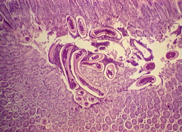
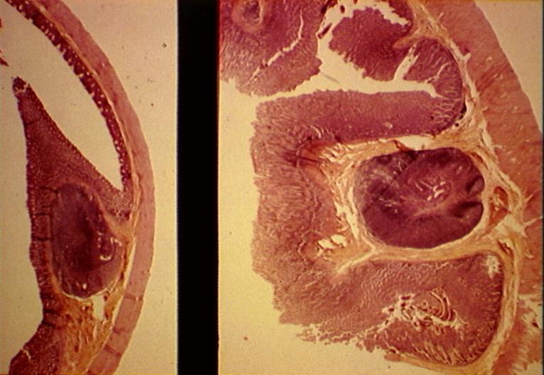
| 19:38, 14 August 2007 | Trichuris vulpis caecum.jpg (file) |  |
100 KB | Caecum showing hyperplastic mucosa and sections of Trichuris vulpis (dog whipworm) and eggs . | 1 |
| 19:39, 14 August 2007 | Trichuris vulpis caecum comparative.jpg (file) |  |
69 KB | Dog caecum: left normal, right with mucosal hyperplasia and inflammation caused by Trichuris vulpis . | 1 |
| 19:45, 14 August 2007 | Trichuris ovis.jpg (file) |  |
55 KB | Trichuris ovis in caecum. | 1 |
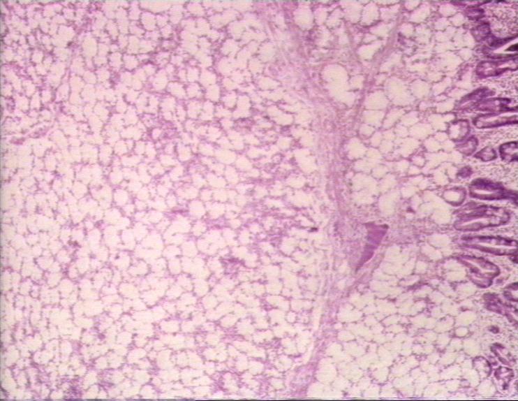
| 20:17, 14 August 2007 | Johnes disease histological.jpg (file) |  |
100 KB | Johne's disease in the ileum- histological. | 1 |
| 20:19, 14 August 2007 | Johnes disease proliferative ileitis.jpg (file) |  |
63 KB | Ileum opened: proliferative ileitis in Johne's disease. | 1 |
| 20:24, 14 August 2007 | Porcine intestinal adenomatosis campylobacter.jpg (file) |  |
107 KB | Section of ileum stained to reveal Campylobacter mucosalis in intracellular porcine intestinal adenomatosis . | 1 |
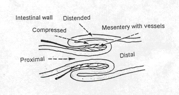
| 09:14, 15 August 2007 | Intussusception.jpg (file) |  |
34 KB | A diagrammatic representation of intussusception. | 1 |
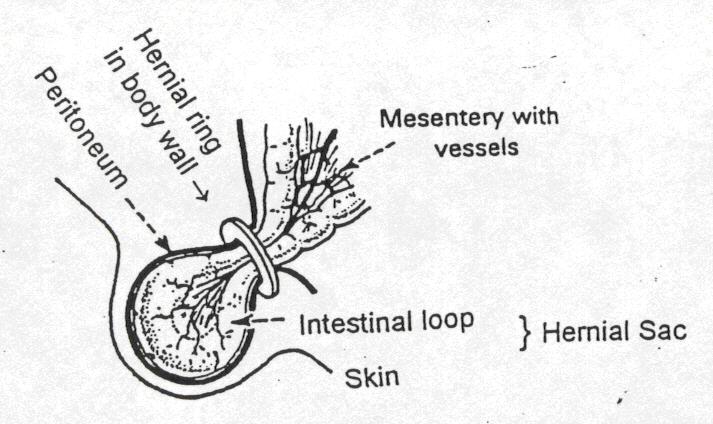
| 09:15, 15 August 2007 | Hernial sac.jpg (file) |  |
51 KB | A diagrammatic representation of the hernial sac. | 1 |
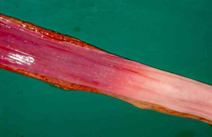
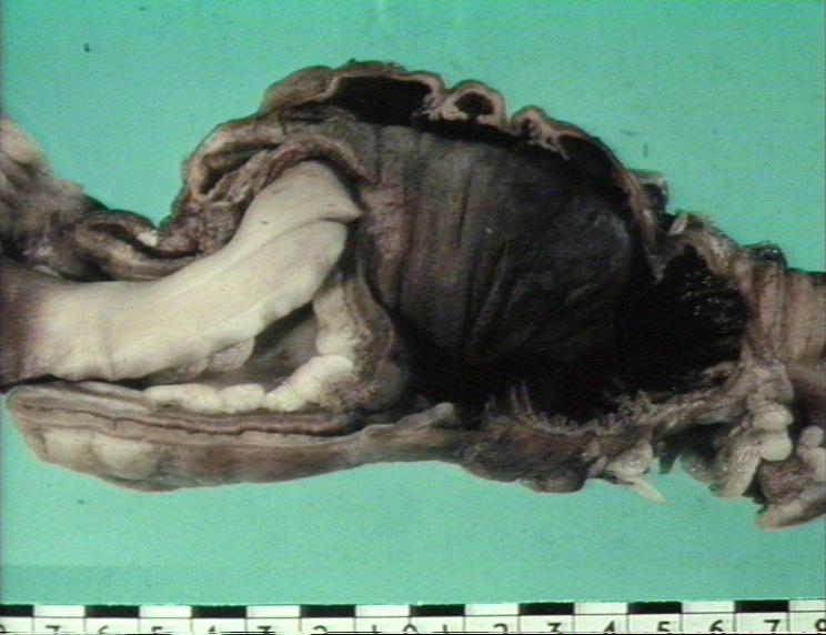
| 09:18, 15 August 2007 | Stomach diaphragmatic hernia.jpg (file) |  |
13 KB | Diaphragmatic hernia: stomach displaced into the thorax, lungs compressed and diaphragm displaced caudally. | 1 |
| 09:20, 15 August 2007 | Volvulus.jpg (file) |  |
54 KB | Volvulus. | 1 |
| 09:23, 15 August 2007 | Intussuceptionphoto.jpg (file) |  |
56 KB | Intussusception. | 1 |
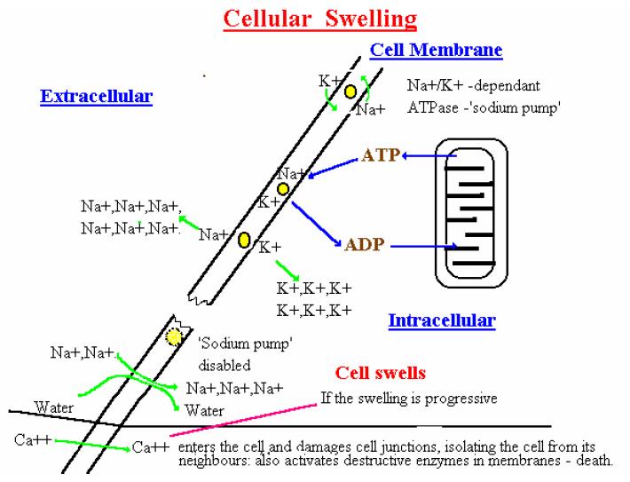
| 18:22, 20 August 2007 | Cellular swelling diagram.jpg (file) |  |
56 KB | The mechanism of cellular swelling. Pending permission from Brian Smyth. | 1 |
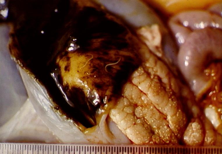
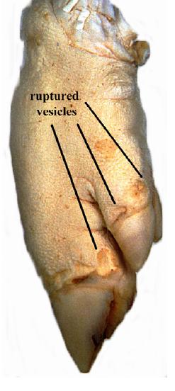
| 18:27, 20 August 2007 | Hydropic degeneration foot and mouth pig foot.jpg (file) |  |
23 KB | Pig foot affected by Foot and Mouth Disease. Shows ruptures vesicles (Ballooning degeneration). Pending permission from Brian Smyth. | 1 |
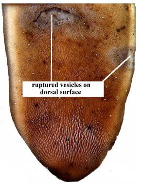
| 18:30, 20 August 2007 | Hydropic degneration foot and mouth ox tongue.jpg (file) |  |
25 KB | The ox tongue in Foot and Mouth Disease. Ruptured vesicles (ballooning degeneration) are seen on the dorsal surface. Pending permission from Brian Smyth. | 1 |
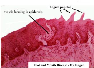
| 18:31, 20 August 2007 | Hydropic degeneration foot and mouth ox tongue histo 1.jpg (file) |  |
16 KB | Histological appearance of ox tongue in Foot and Mouth Disease. A vesicle is forming in the epidermis. Pending permission from Brian Smyth. | 1 |
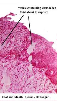
| 18:32, 20 August 2007 | Hydropic degeneration foot and mouth ox tongue histo 2.jpg (file) |  |
17 KB | Histological appearance of ox tongue in Foot and Mouth Disease. A virus-containing vesicle in the epidermis is close to rupture. Pending permission from Brian Smyth. | 1 |
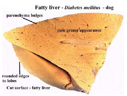
| 18:47, 20 August 2007 | Fatty liver.jpg (file) |  |
21 KB | The gross appearance of a liver showing fatty change. Pending permission from Brian Smyth. | 1 |
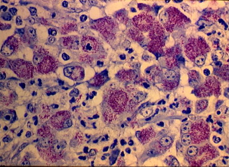
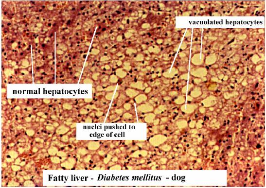
| 18:54, 20 August 2007 | Fatty liver histo.jpg (file) |  |
68 KB | Histological appearance of a liver showing fatty change. Fat vacuoles in the cytoplasm displace the nuclei to the periphery of the cells. Pending permission from Brian Smyth. | 1 |
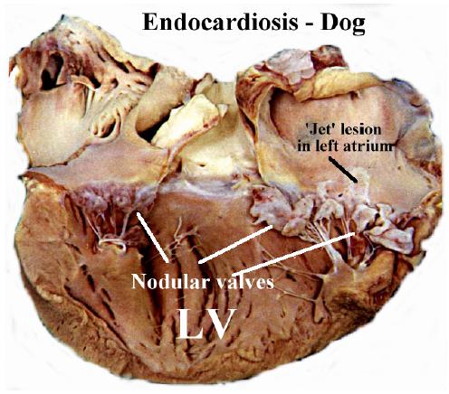
| 18:56, 20 August 2007 | Endocardiosis.jpg (file) |  |
45 KB | Gross appearance of endocardiosis in the dog. Pending permission from Brian Smyth. | 1 |
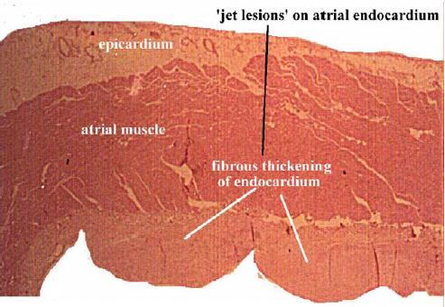
| 18:57, 20 August 2007 | Endocardiosis histo 2.jpg (file) |  |
35 KB | Histological appearance of endocardiosis in the dog. | 1 |
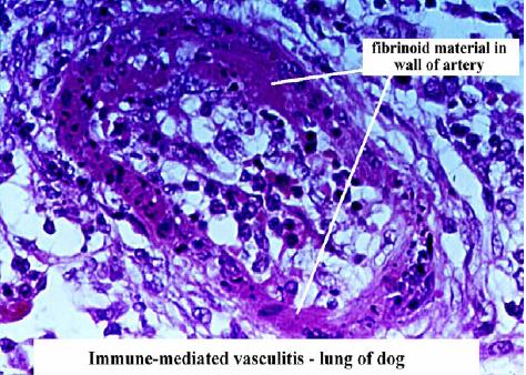
| 19:00, 20 August 2007 | Fibrinoid degeneration immune mediated vasculitis.jpg (file) |  |
54 KB | An example of fibrinoid degeneration in immune mediated vasculitis in the lung of the dog. Pending permission from Brain Smyth. | 1 |
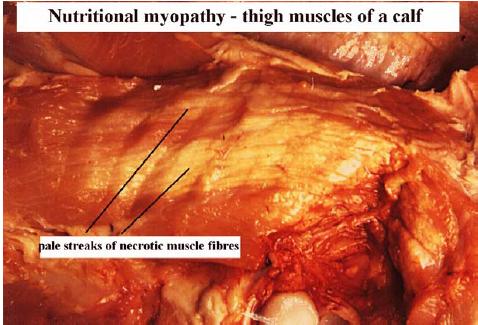
| 19:06, 20 August 2007 | Nutritional myopathy.jpg (file) |  |
34 KB | Gross appearance of hyaline degeneration of skeletal muscle due to vitamin E/ selenium deficiency. Pending permission from Brain Smyth. | 1 |
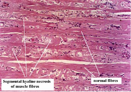
| 19:06, 20 August 2007 | Nutritional myopathy histo.jpg (file) |  |
39 KB | Histological appearance of hyaline degeneration of skeletal muscle due to vitamin E/ selenium deficiency. Pending permission from Brain Smyth. | 1 |
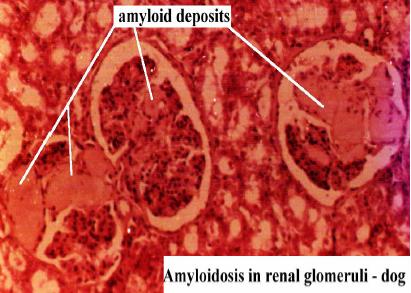
| 19:08, 20 August 2007 | Amyloidosis.jpg (file) |  |
29 KB | Histological appearance of amyloidosis. Pending permission from Brian Smyth. | 1 |
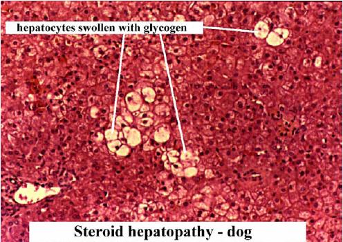
| 19:10, 20 August 2007 | Glycogen infiltration.jpg (file) |  |
53 KB | Glycogen infiltration due to steroid hepatopathy in the dog. Pending permission from Brian Smyth. | 1 |
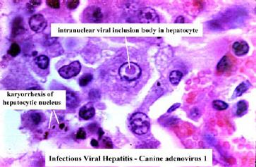
| 19:24, 20 August 2007 | Viral inclusion canine adenovirus 1.jpg (file) |  |
23 KB | Viral inclusion due to canine adenovirus 1. Pending permission from Brain Smyth. | 1 |
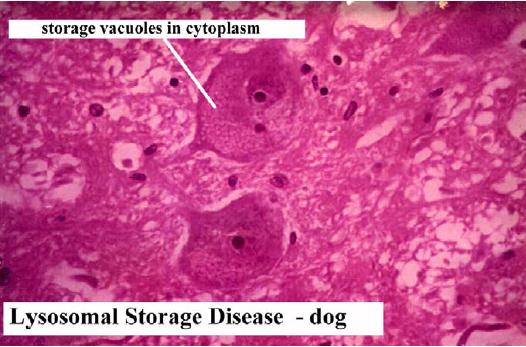
| 19:41, 20 August 2007 | Lysosomal storage disease.jpg (file) |  |
41 KB | Histological appearance of lysomsomal storage disease in the dog. Pending permission from Brian Smyth. | 1 |
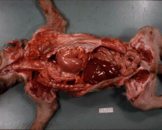
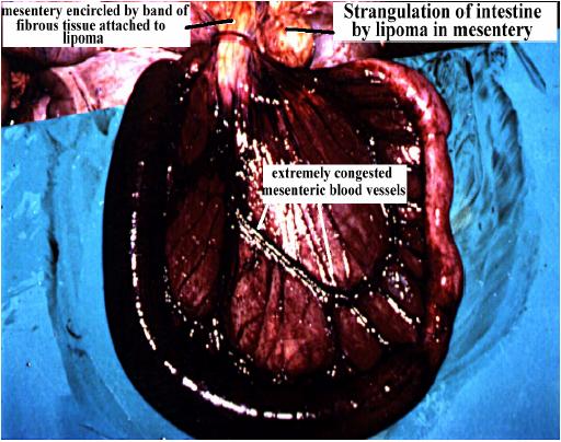
| 20:33, 20 August 2007 | Strangulation of intestine.jpg (file) |  |
49 KB | Stangulation of the intestine by lipoma in the mesentery. Pending permsission from Brian Smyth. | 1 |
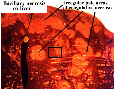
| 20:51, 20 August 2007 | Coagulative necrosis bacillary necrosis.jpg (file) |  |
35 KB | Coagulative necrosis due to bacillary necrosis. Pending permission form Brian Smyth. | 1 |
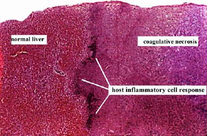
| 20:52, 20 August 2007 | Coagulative necrosis histo.jpg (file) |  |
35 KB | Histological appearance of coagulative necrosis. Pending permission from Brian Smyth. | 1 |