Uploads by Lizzies
Jump to navigation
Jump to search
This special page shows all uploaded files.
| Date | Name | Thumbnail | Size | Description | Versions |
|---|---|---|---|---|---|
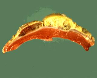
| 18:52, 9 August 2007 | Traumatic pericarditis.jpg (file) |  |
7 KB | 1 | |
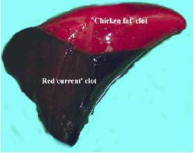
| 09:51, 21 August 2007 | Clot.jpg (file) |  |
8 KB | A post-mortem clot, showing the upper "chicken-fat" clot and the lower "redcurrant-jelly" clot. Pending permission from Brian Smyth. | 1 |
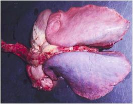
| 09:58, 21 August 2007 | Hypostatic congestion.jpg (file) |  |
11 KB | Hypostatic congestion in the lungs. Pending permission from Brian Smyth. | 1 |
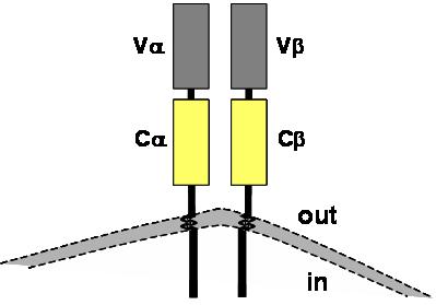
| 16:10, 4 September 2007 | TcR structure.jpg (file) |  |
11 KB | Diagram showing the basic structure of a TcR. Pending permission from John Hopkins. | 1 |
| 16:30, 29 August 2007 | Anasarca.jpg (file) |  |
12 KB | Anasarca. Pending permission from Brian Smyth. | 1 |
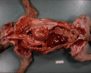
| 09:18, 15 August 2007 | Stomach diaphragmatic hernia.jpg (file) |  |
13 KB | Diaphragmatic hernia: stomach displaced into the thorax, lungs compressed and diaphragm displaced caudally. | 1 |
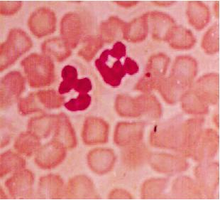
| 09:28, 31 August 2007 | Neutrophils.jpg (file) |  |
13 KB | Neutrophils (H&E). Pending permission from Brian Smyth. | 1 |
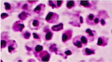
| 09:34, 31 August 2007 | Macrophages.jpg (file) |  |
15 KB | Macrophages. Pending permission from Brian Smyth. | 1 |
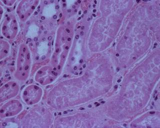
| 21:16, 13 August 2007 | Pulpy kidney disease.jpg (file) |  |
15 KB | Abrupt transition between viable and necrotic renal tubules in pulpy kidney disease. | 1 |
| 10:31, 7 September 2007 | Consolidation and haemorrhage lung.jpg (file) |  |
16 KB | Consolidation and haemorrhage in the lung. Courtesy of BioMed Archive. | 1 |
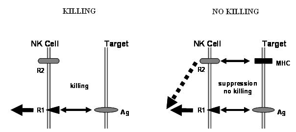
| 16:27, 4 September 2007 | NK cell killing.jpg (file) |  |
16 KB | Diagrammatic representation of the receptors involved in NK cell killing. Pending permission from John Hopkins. | 1 |
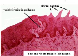
| 18:31, 20 August 2007 | Hydropic degeneration foot and mouth ox tongue histo 1.jpg (file) |  |
16 KB | Histological appearance of ox tongue in Foot and Mouth Disease. A vesicle is forming in the epidermis. Pending permission from Brian Smyth. | 1 |
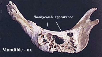
| 09:43, 31 August 2007 | Lumpy jaw mandible.jpg (file) |  |
17 KB | "Honeycomb" appearance of mandible, following actinomycosis (Lumpy Jaw). Pending permission from Brian Smyth. | 1 |
| 09:28, 31 August 2007 | Eosinophils.jpg (file) |  |
17 KB | Eosinophils (H&E). Pending permission from Brian Smyth. | 1 |
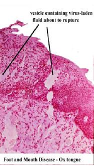
| 18:32, 20 August 2007 | Hydropic degeneration foot and mouth ox tongue histo 2.jpg (file) |  |
17 KB | Histological appearance of ox tongue in Foot and Mouth Disease. A virus-containing vesicle in the epidermis is close to rupture. Pending permission from Brian Smyth. | 1 |
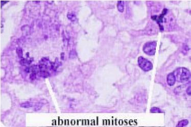
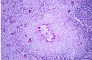
| 11:42, 3 September 2007 | Abnormal mitoses.jpg (file) |  |
17 KB | Abnormal mitoses. Pending permission from Brian Smyth. | 1 |
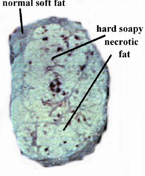
| 21:15, 20 August 2007 | Fat necrosis ox subcutis.jpg (file) |  |
18 KB | Gross appearance of fat necrosis in the ox subcutis. Pending permission from Brian Smyth. | 1 |
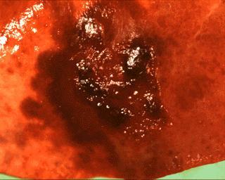
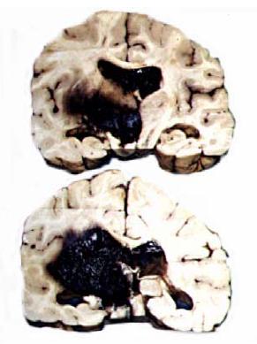
| 16:34, 29 August 2007 | Haemorrhage in the brain.jpg (file) |  |
19 KB | Haemorrhage in the brain. Pending permission from Brian Smyth. | 1 |
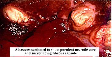
| 21:05, 20 August 2007 | Abscess centre and capsule.jpg (file) |  |
19 KB | Gross appearance of an abscess, showing the necrotic centre and the capsule. Pending permission from Brian Smyth. | 1 |
| 21:27, 20 August 2007 | Calcification.jpg (file) |  |
19 KB | Histological appearance of calcification. Pending permission from Brian Smyth. | 1 |
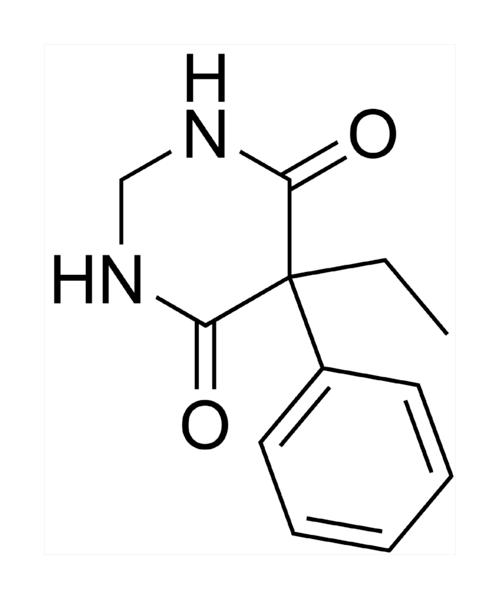
| 17:09, 25 August 2010 | Primidone.jpg (file) |  |
19 KB | {{Information |Description=The structure of primidone. |Source=Wikimedia Commons |Date=Originally uploaded 9th February 2006. Uploaded to WikiVet 25th August 2010 |Author=Jesse |Permission=This image has been released to the public domain by the author. | | 1 |
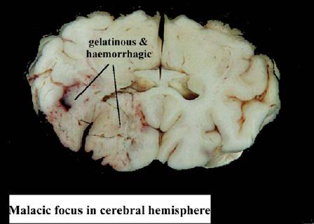
| 20:53, 20 August 2007 | Malacia.jpg (file) |  |
19 KB | 1 | |
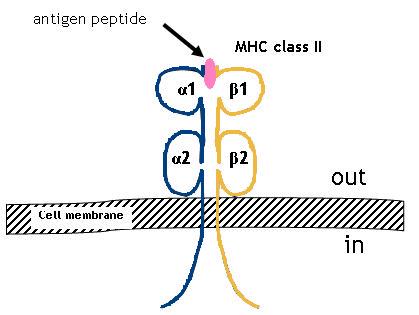
| 09:57, 5 September 2007 | MHC II.jpg (file) |  |
19 KB | Structure of MHC II. Pending permission from John Hopkins. | 1 |
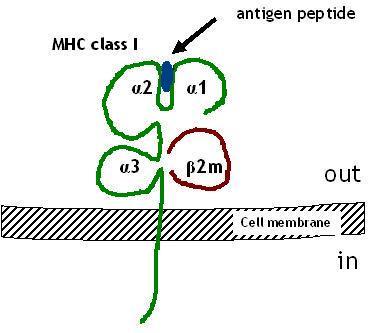
| 09:29, 5 September 2007 | MHC I.jpg (file) |  |
19 KB | Structure of MHC class I. Pending permission from John Hopkins. | 1 |
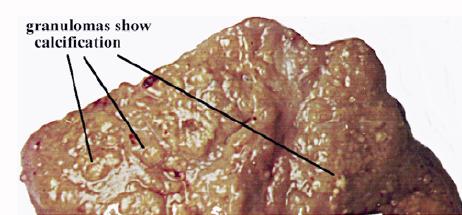
| 09:48, 31 August 2007 | Tuberculous granulomas.jpg (file) |  |
20 KB | Gross appearance of tuberculous granulomas in the lymph node of the ox. These granulomas show a degree of calcification. Pending permission fromo Brian Smyth. | 1 |
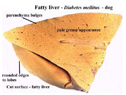
| 18:47, 20 August 2007 | Fatty liver.jpg (file) |  |
21 KB | The gross appearance of a liver showing fatty change. Pending permission from Brian Smyth. | 1 |
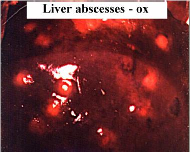
| 21:01, 20 August 2007 | Liver abscess.jpg (file) |  |
21 KB | Abscesses in the liver. Pending permission from Brian Smyth. | 1 |
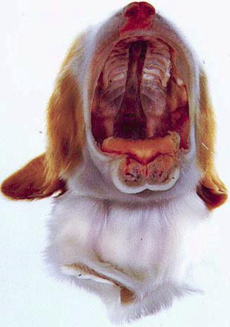
| 15:27, 3 September 2007 | Cleft palate.jpg (file) |  |
21 KB | Cleft palate in the cat. Pending permission from Brian Smyth. | 1 |
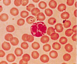
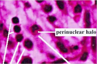
| 09:32, 31 August 2007 | Lymphocytes and plasma cell.jpg (file) |  |
21 KB | Note the 4 lymphocytes on the right hand side, and the plasma cell with the perinuclear halo. Pending permission from Brian Smyth. | 1 |
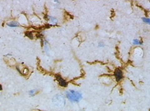
| 11:38, 27 March 2008 | Microglia.jpg (file) |  |
21 KB | Microglia cells stained immunohisotchemically for lectins. Image sourced from [http://commons.wikimedia.org/wiki/Image:Mikroglej_1.jpg|WikiMedia Commons], where it is attributed to Grzegorz Wicher. | 1 |
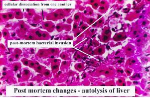
| 10:35, 21 August 2007 | Autolysis of liver.jpg (file) |  |
21 KB | Histological appearance of autolysis in the liver. Pending permission from Brian Smyth. | 1 |
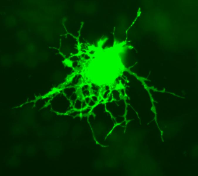
| 10:48, 27 March 2008 | Oligodendrocyte.jpg (file) |  |
22 KB | Oligodendrocyte. Image obtained from [http://commons.wikimedia.org/wiki/Image:Oligodendrocyte.png WikiMedia Commons]. | 1 |
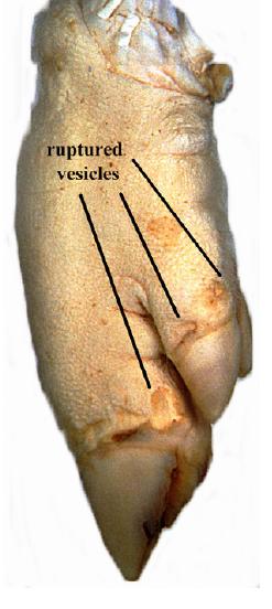
| 18:27, 20 August 2007 | Hydropic degeneration foot and mouth pig foot.jpg (file) |  |
23 KB | Pig foot affected by Foot and Mouth Disease. Shows ruptures vesicles (Ballooning degeneration). Pending permission from Brian Smyth. | 1 |
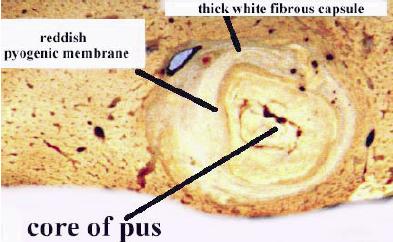
| 09:23, 31 August 2007 | Acute abscess.jpg (file) |  |
23 KB | Cross section through an acute abscess. Pending permission from Brian Smyth. | 1 |
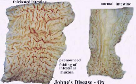
| 10:25, 31 August 2007 | Johnes disease comparative.jpg (file) |  |
23 KB | Comparative gross appearance of Johne's Disease in the ox. The intestine on the right is diseased, whereas that on the left is normal. Pending permission from Brian Smyth. | 1 |
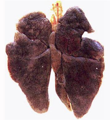
| 12:26, 21 August 2007 | Anthracosis gross.jpg (file) |  |
23 KB | Gross appearance of antrhacosis in the lungs of a dog. Pending permission from Brian Smyth. | 1 |
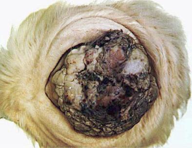
| 13:17, 3 September 2007 | Squamous cell carcinoma eye.jpg (file) |  |
23 KB | Squamous cell carcinoma of the eye of a hereford cow. UV light contributes to causing the tumour. Pending permission from Brian Smyth. | 1 |
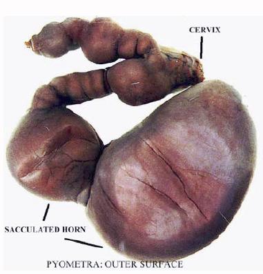
| 10:09, 31 August 2007 | Pyometra.jpg (file) |  |
23 KB | Pyometra - outside surface of the uterus. Pending permission from Brian Smyth. | 1 |
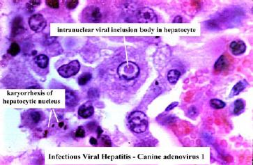
| 19:24, 20 August 2007 | Viral inclusion canine adenovirus 1.jpg (file) |  |
23 KB | Viral inclusion due to canine adenovirus 1. Pending permission from Brain Smyth. | 1 |
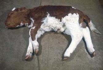
| 16:28, 7 August 2007 | Cow2.jpg (file) |  |
24 KB | 1 | |
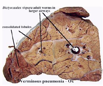
| 10:20, 31 August 2007 | Lungworm.jpg (file) |  |
24 KB | ''Dictyocaulus vivipara'' in the lung of the ox. Causes verminous pneumonia. Pending permission from Brian Smyth. | 1 |
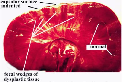
| 12:18, 31 August 2007 | Renal dysplasia dog gross.jpg (file) |  |
24 KB | Gross apperarance of renal dysplasia in the dog. Pending permission from Brian Smyth. | 1 |
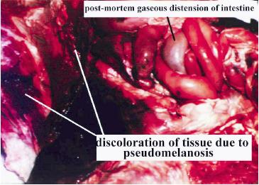
| 12:01, 21 August 2007 | Pseudomelanosis.jpg (file) |  |
24 KB | Appearance of pseudomelanosis. Pending permission from Brian Smyth. | 1 |
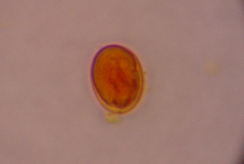
| 16:17, 11 August 2010 | Giardia Cyst.jpg (file) |  |
25 KB | {{Information |Description= Giardia cyst from a dog |Source= Wikimedia Commons |Date= 6 September 2006 |Author= Joel Mills |Permission= Creative Commons Attribution-Share Alike 3.0 Unported license }} | 1 |
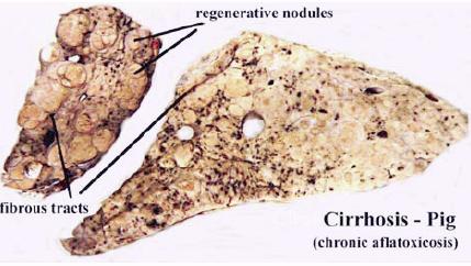
| 10:31, 31 August 2007 | Cirrhosis pig.jpg (file) |  |
25 KB | Cirrhosis, due to chronic liver damage in the pig. Pending permission fro Brian Smyth. | 1 |
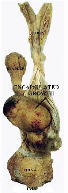
| 11:14, 3 September 2007 | Benign leiomyoma.jpg (file) |  |
25 KB | Benign neoplasm (leiomyoma). Note that the growth is encapsulated. Pending permission from Brian Smyth. | 1 |
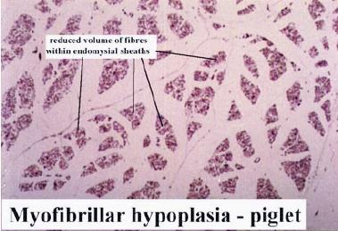
| 11:50, 31 August 2007 | Myofibrillar hypoplasia.jpg (file) |  |
25 KB | Myofibrillar hypoplasia in the piglet. Pending permission from Brian Smyth. | 1 |
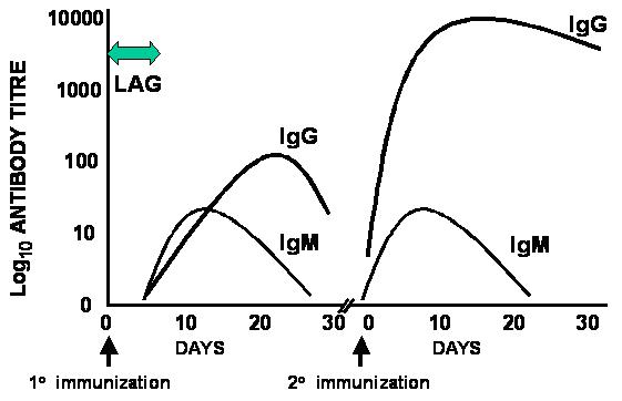
| 15:46, 4 September 2007 | Primary and secondary response.jpg (file) |  |
25 KB | Time course of the primary and secondary responses. Pending permission from John Hopkins. | 1 |
| 18:30, 20 August 2007 | Hydropic degneration foot and mouth ox tongue.jpg (file) |  |
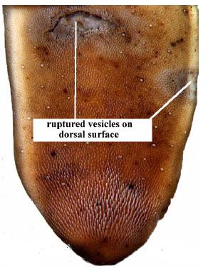
25 KB | The ox tongue in Foot and Mouth Disease. Ruptured vesicles (ballooning degeneration) are seen on the dorsal surface. Pending permission from Brian Smyth. | 1 |
| 16:17, 4 January 2008 | Secondaryhyperparathyroidism.jpg (file) |  |
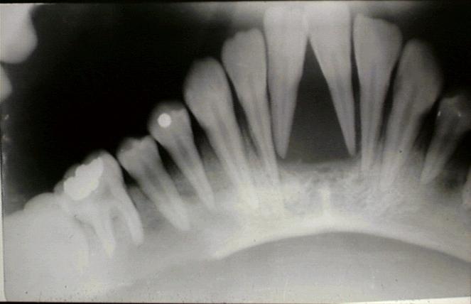
25 KB | Dental radiograph of secondary hyperparathyroidism. Note the demineralisation of bone around the tooth roots. Courtesy of Biomed Image Archive. | 1 |