Uploads by Nabrown
This special page shows all uploaded files.
| Date | Name | Thumbnail | Size | Description | Versions |
|---|---|---|---|---|---|
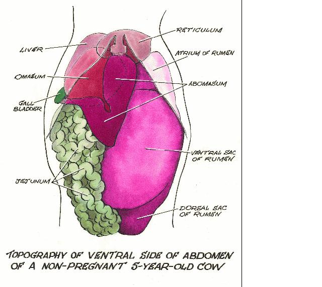
| 11:31, 31 July 2008 | Abdomen development older cow.jpg (file) |  |
56 KB | Copyright Prof. Pat Mccarthy | 1 |
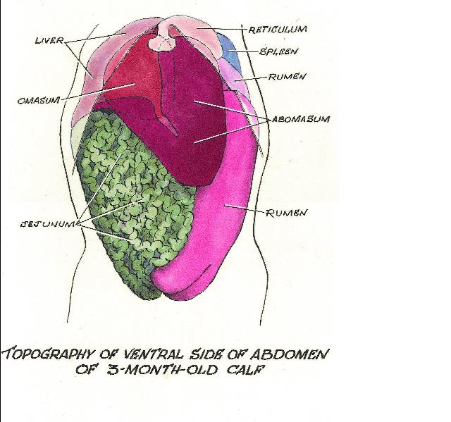
| 11:31, 31 July 2008 | Abdomen development young cow.jpg (file) |  |
57 KB | Copyright Prof. Pat Mccarthy | 1 |
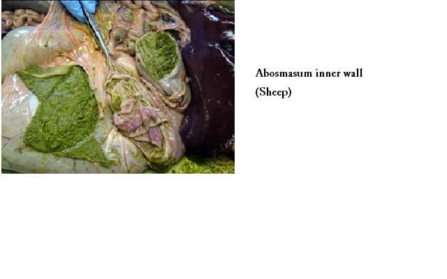
| 17:53, 8 July 2008 | Abomasum Anatomy Sheep.jpg (file) |  |
27 KB | Copywright RVC 2008 | 1 |
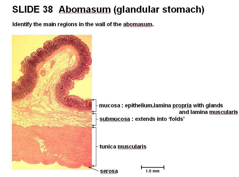
| 17:54, 8 July 2008 | Abomasum Histology Sheep.jpg (file) |  |
64 KB | Copywright RVC 2008 | 1 |
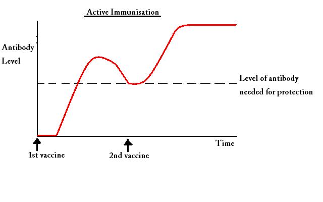
| 16:12, 25 August 2008 | Active Immunisation.jpg (file) |  |
17 KB | Copyright nabrown RVC 2008 | 1 |
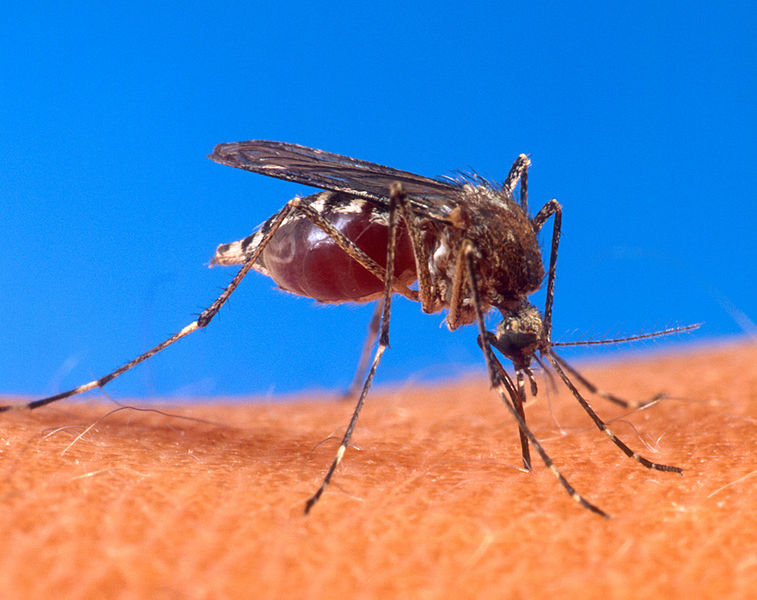
| 20:47, 9 November 2008 | Aedes aegypti.jpg (file) |  |
80 KB | Picture from the USDA website at http://www.ars.usda.gov/is/graphics/photos/aug00/k4705-9.htm Wikimedia Commons | 1 |
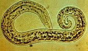
| 11:38, 22 December 2008 | Aeurostrongylus abstrusus.jpg (file) |  |
7 KB | Courtesy of the Laboratory of Parasitology, University of Pennsylvania School of Veterinary Medicine | 1 |
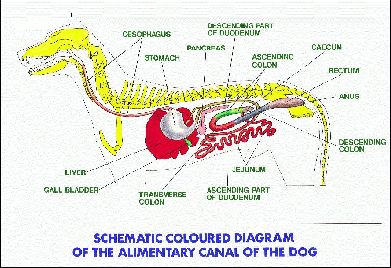
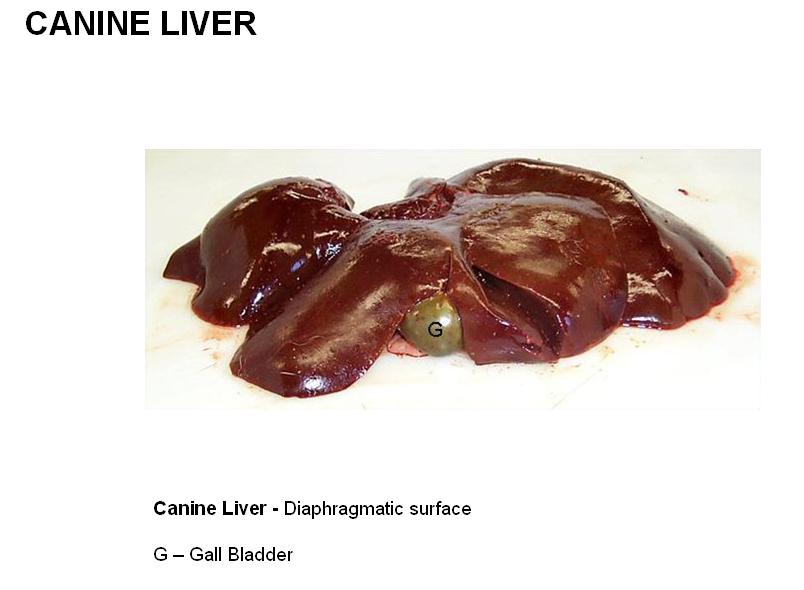
| 11:53, 31 July 2008 | Alimentary Canine.jpg (file) |  |
117 KB | Copyright Prof. Pat Mccarthy | 1 |
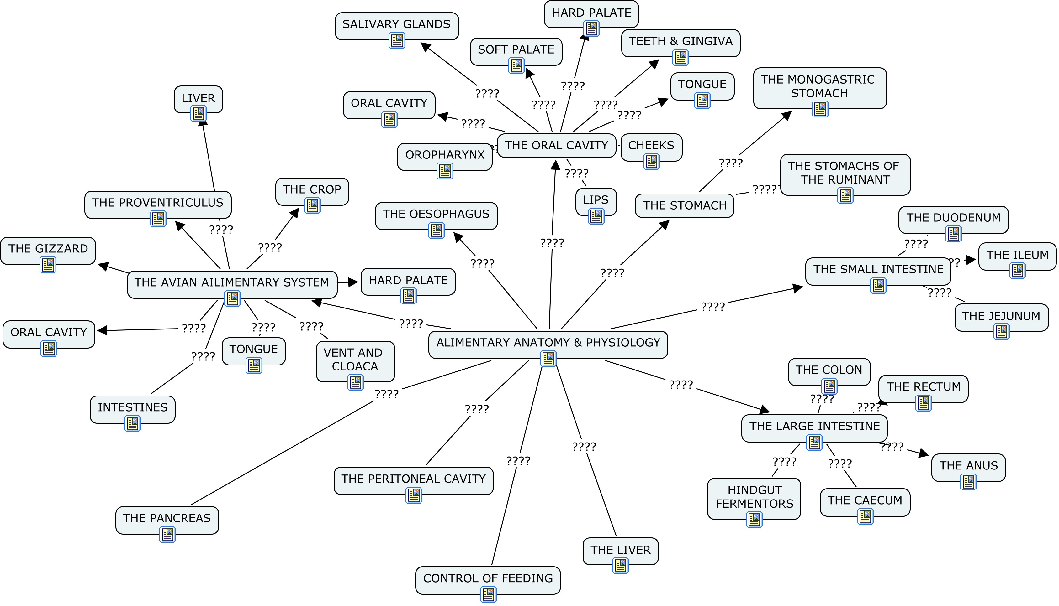
| 12:53, 18 July 2008 | Alimentary System Concept Map - Anatomy & Physiology.jpg (file) |  |
613 KB | Copyright nabrown RVC 2008 | 1 |
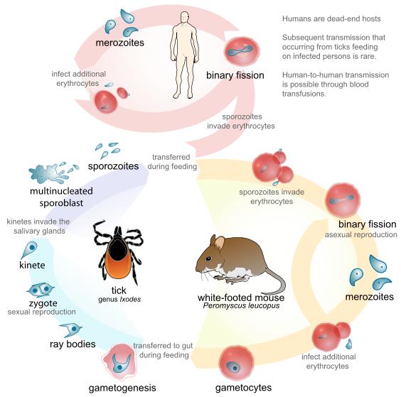
| 17:32, 22 November 2008 | Alternative Babesia life cycle diagram.jpg (file) |  |
42 KB | Mariana Ruiz Villarreal Life cycle of the Parasite Babesia, (B.microti or B.divergens) including the infection to humans http://en.wikipedia.org/wiki/Image:Babesia_life_cycle_human_en.svg Wikimedia Commons | 1 |
| 16:54, 14 July 2008 | Anatomy of the Avian Liver.jpg (file) |  |
49 KB | Copywright RVC 2008 | 1 |
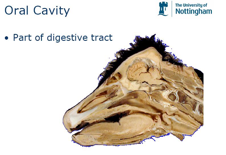
| 10:36, 29 July 2008 | Anatomy of the Oral Cavity.jpg (file) |  |
54 KB | Copyright Nottingham 2008 | 1 |
| 13:58, 23 December 2008 | Ancylostoma.jpg (file) |  |
5 KB | Courtesy of the Laboratory of Parasitology, University of Pennsylvania School of Veterinary Medicine | 1 |
| 14:36, 22 December 2008 | Anoplocephala.jpg (file) |  |
5 KB | Joaquim Castellà Veterinary Parasitology Universitat Autònoma de Barcelona | 1 |
| 17:59, 24 October 2008 | Arthropod classifiation.jpg (file) |  |
17 KB | Copyright nabrown RVC | 1 |
| 13:01, 31 July 2008 | Arytenoid cartilages.jpg (file) |  |
20 KB | Copyright RVC 2008 | 1 |
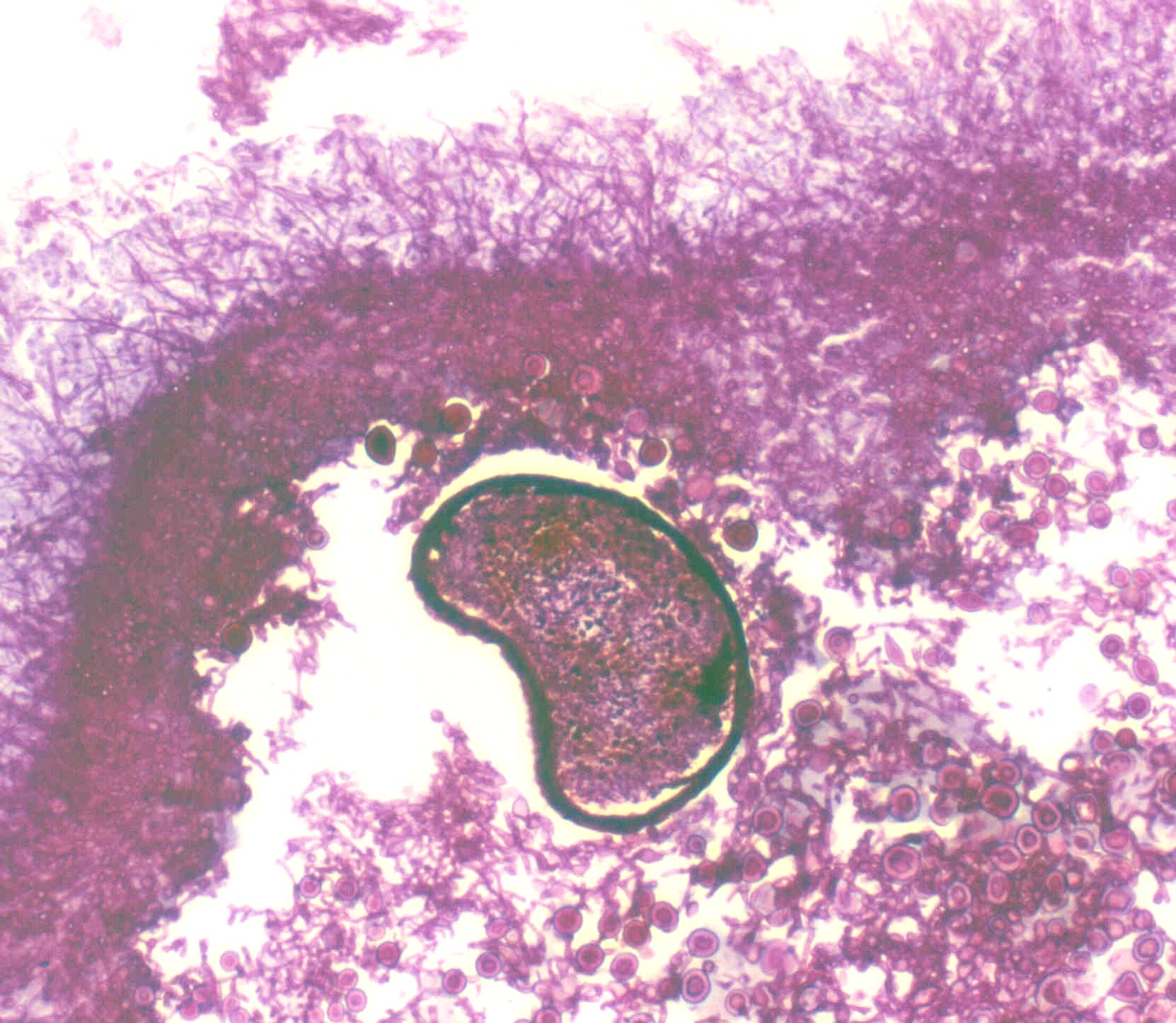
| 16:35, 4 June 2009 | Aspergillus cleistothecia.jpg (file) |  |
344 KB | Aspergillus cleistothecia perithecium showing Hulle cells which are the sexual form (ascospores). Sampled from an equine gutteral pouch mycosis lesion and stained with Gram stain. Copyright - Professor Andrew N. Rycroft, BSc, PHD, C. Biol.F.I.Biol., FRC | 1 |
| 16:30, 4 June 2009 | Aspergillus in vivo.jpg (file) |  |
48 KB | Copyright Professor Andrew N. Rycroft, BSc, PHD, C. Biol.F.I.Biol., FRCPath | 1 |
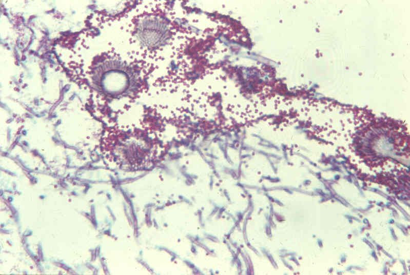
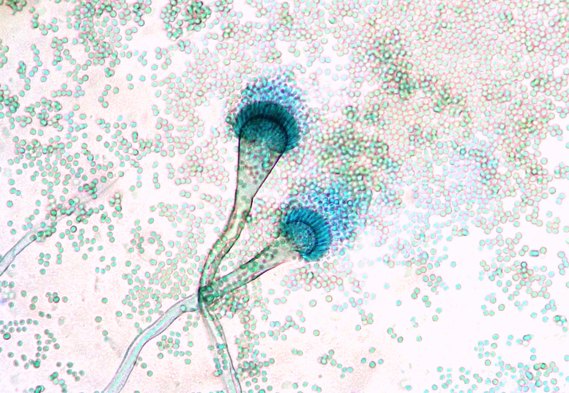
| 16:32, 4 June 2009 | Aspergillus sporing heads.jpg (file) |  |
521 KB | Aspergillus sporing heads Copyright - Professor Andrew N. Rycroft, BSc, PHD, C. Biol.F.I.Biol., FRCPath | 1 |
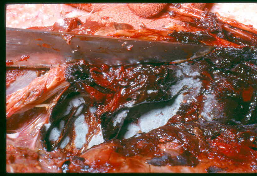
| 16:28, 4 June 2009 | Aspergillus swan.jpg (file) |  |
69 KB | Aspergillus in a swan. It is usually found near an interface where gas exchange occurs. For example, the air sacs. Copyright - Professor Andrew N. Rycroft, BSc, PHD, C. Biol.F.I.Biol., FRCPath | 1 |
| 15:51, 16 July 2008 | Avian Cloaca Diagram.jpg (file) |  |
71 KB | Copyright RVC 2008 | 1 |
| 14:59, 16 July 2008 | Avian GIT.jpg (file) |  |
36 KB | Copyright Nottingham 2008 | 1 |
| 12:11, 29 July 2008 | Avian Tongue and Oral Cavity.jpg (file) |  |
67 KB | Copyright RVC 2008 | 1 |
| 17:17, 22 November 2008 | Babesia Life Cycle.jpg (file) |  |
30 KB | Babesia Life Cycle Diagram - Dennis Jacobs & Mark Fox RVC | 1 |
| 19:08, 23 November 2008 | Balantidium pig.jpg (file) |  |
9 KB | ''Balantidium'' from a pig - Joaquim Castellà Veterinary Parasitology Universitat Autònoma de Barcelona | 1 |
| 19:08, 23 November 2008 | Balantidium pig trophozoite.jpg (file) |  |
68 KB | ''Balantidium'' trophozoite from a pig - Joaquim Castellà Veterinary Parasitology Universitat Autònoma de Barcelona | 1 |
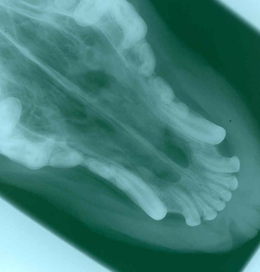
| 11:17, 30 July 2008 | Bird skull.jpg (file) |  |
36 KB | Copyright RVC 2008 | 1 |
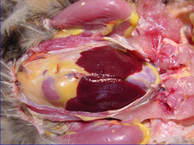
| 19:05, 23 November 2008 | Blackhead lesions in turkey liver.jpg (file) |  |
20 KB | Histomoniasis, or Blackhead disease. Large, pale areas in the liver of a bird infected with Histomonas sp. Photo credit:Milton Friend http://en.wikipedia.org/wiki/Image:Histomoniasis.jpg | 1 |
| 15:16, 10 July 2008 | Blood Flow In and Away from the Liver.jpg (file) |  |
17 KB | Copyright nabrown RVC 2008 | 1 |
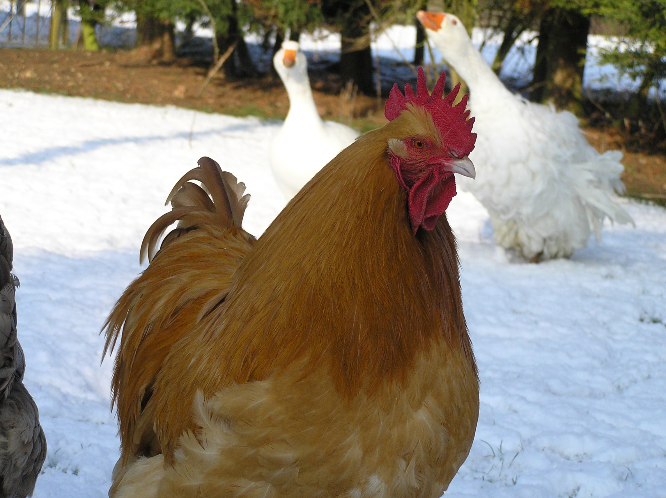
| 10:09, 16 November 2008 | Buff orpington.jpg (file) |  |
740 KB | Copyright nabrown RVC | 1 |
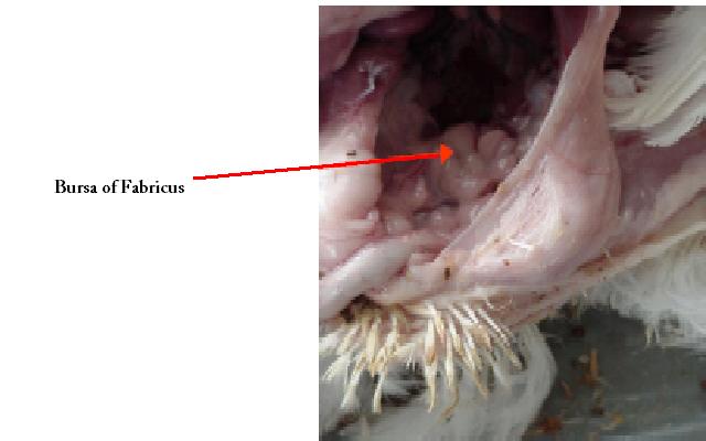
| 15:23, 16 July 2008 | Bursa of Fabricus.jpg (file) |  |
23 KB | Copyright Nottingham 2008 | 1 |
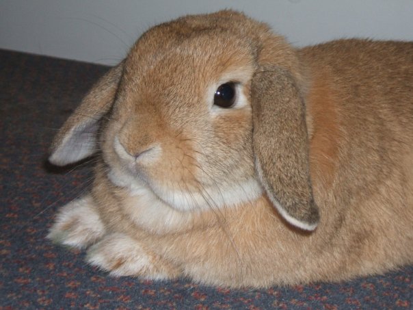
| 10:05, 11 September 2008 | Buzz bunny.jpg (file) |  |
55 KB | Copyright L. Drew RVC | 1 |
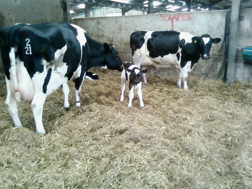
| 18:00, 23 November 2008 | Calf.jpg (file) |  |
235 KB | nabrown RVC Daisy! | 1 |
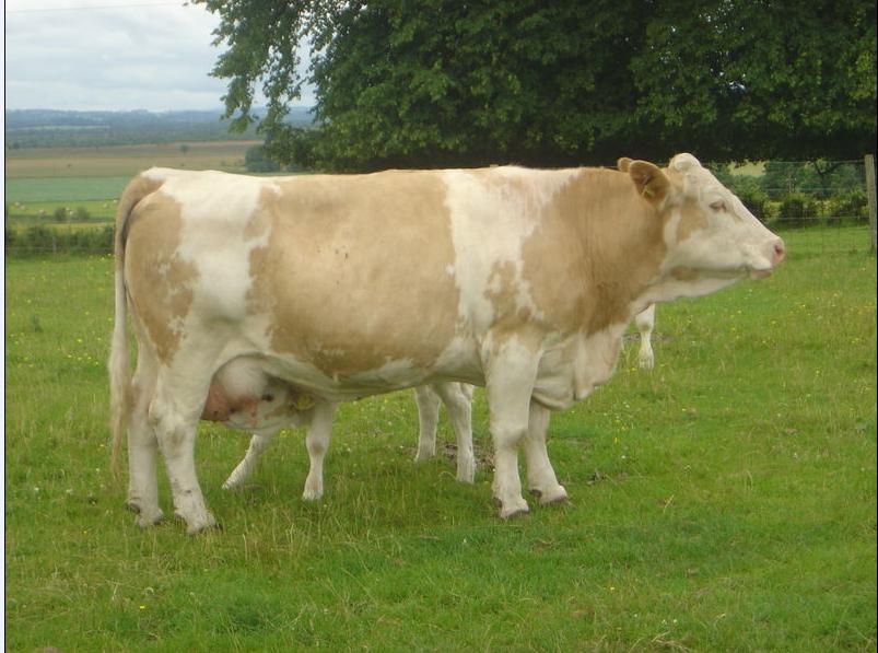
| 11:46, 31 July 2008 | Calf Suckling.jpg (file) |  |
71 KB | Copyright David Monniaux | 1 |
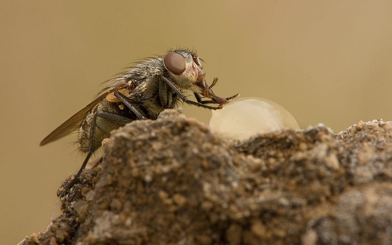
| 20:32, 9 November 2008 | Calliphoridae.jpg (file) |  |
49 KB | Pollenia rudis (Calliphoridae) - Richard Bartz, Munich - Wikimedia Commons | 1 |
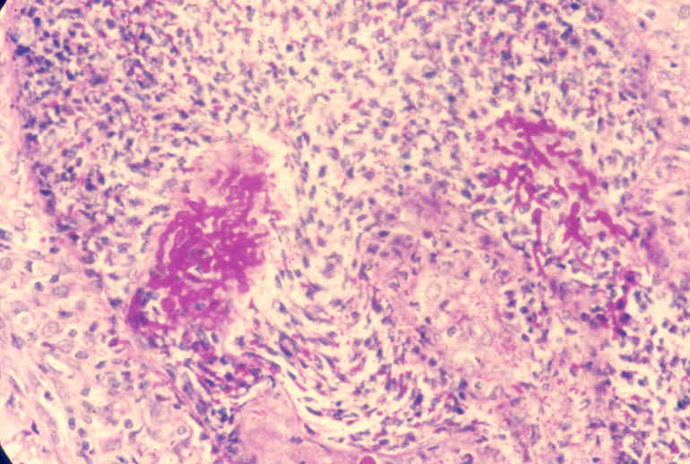
| 16:40, 4 June 2009 | Candida.jpg (file) |  |
549 KB | Copyright - Professor Andrew N. Rycroft, BSc, PHD, C. Biol.F.I.Biol., FRCPath | 1 |
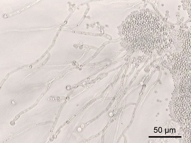
| 15:55, 23 April 2009 | Candida albicans.jpg (file) |  |
82 KB | http://commons.wikimedia.org/wiki/File:Candida_albicans.jpg Microscopic image (200-fold magnification) of Candida albicans ATCC 10231, grown on cornmeal agar medium with 1% Tween80. Wikimedia Commons | 1 |
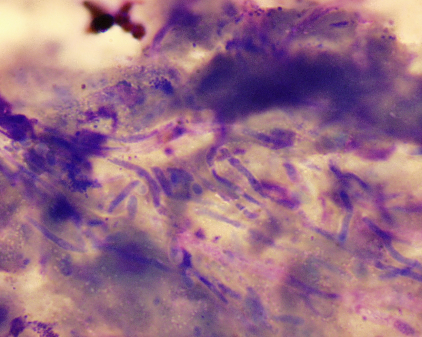
| 16:41, 4 June 2009 | Candida in vivo.jpg (file) |  |
58 KB | Pseudomycelium in vivo Copyright - Professor Andrew N. Rycroft, BSc, PHD, C. Biol.F.I.Biol., FRCPath | 1 |
| 14:32, 10 July 2008 | Canine Gallbladder Anatomy.jpg (file) |  |
39 KB | Copyright RVC 2008 | 1 |
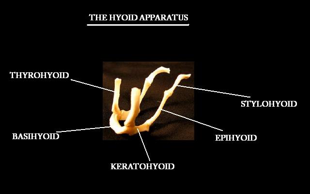
| 12:07, 30 July 2008 | Canine Hyoid.jpg (file) |  |
22 KB | Copyright Nottingham 2008 | 1 |
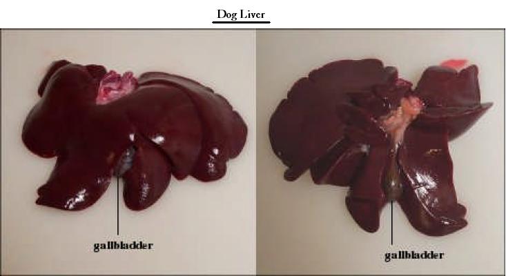
| 09:26, 17 July 2008 | Canine Liver Topography.jpg (file) |  |
27 KB | Copyright Nottingham 2008 | 1 |
| 16:38, 4 June 2009 | Canine nasal asper radiograph.jpg (file) |  |
32 KB | Copyright - Professor Andrew N. Rycroft, BSc, PHD, C. Biol.F.I.Biol., FRCPath | 1 |
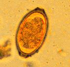
| 18:20, 6 January 2009 | Capilaria.jpg (file) |  |
5 KB | Courtesy of the Laboratory of Parasitology, University of Pennsylvania School of Veterinary Medicine | 1 |
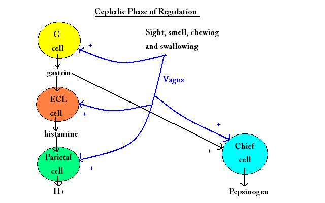
| 09:08, 16 July 2008 | Cephalic phase of secretion diagram.jpg (file) |  |
27 KB | Copyright RVC 2008 | 1 |
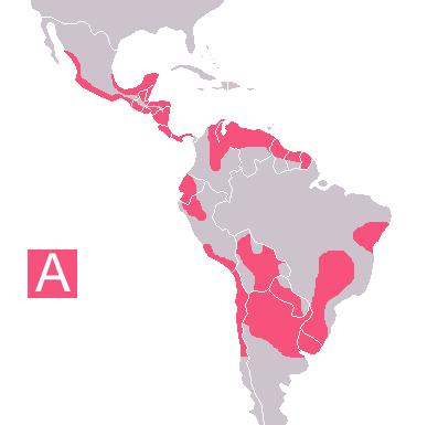
| 17:12, 23 November 2008 | Chagas endemic zones 2005.jpg (file) |  |
12 KB | http://en.wikipedia.org/wiki/Image:Carte_maladie_Chagas.png Wikimedia Commons | 1 |
| 09:30, 15 July 2008 | Choana and Infundibulum Avain.jpg (file) |  |
25 KB | Copyright RVC 2008 | 1 |
| 14:13, 3 July 2008 | Circumvallate Papillae.jpg (file) |  |
79 KB | Copywright RVC 2008 | 1 |
| 19:17, 15 November 2008 | Coccidia.jpg (file) |  |
21 KB | Joel Mills Coccidia oocysts in a fecal flotation from a cat. The cat was underweight and had diarrhea, showing signs of coccidiosis Wikimedia Commons http://en.wikipedia.org/wiki/Image:Coccidia.JPG | 1 |
| 17:06, 17 November 2008 | Coccidia oocyst ruminant.jpg (file) |  |
32 KB | Joaquim Castellà Veterinary Parasitology Universitat Autònoma de Barcelona | 1 |
| 17:06, 17 November 2008 | Coccidia ruminant.jpg (file) |  |
102 KB | Joaquim Castellà Veterinary Parasitology Universitat Autònoma de Barcelona | 1 |