Uploads by Lizzies
Jump to navigation
Jump to search
This special page shows all uploaded files.
| Date | Name | Thumbnail | Size | Description | Versions |
|---|---|---|---|---|---|
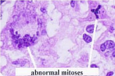
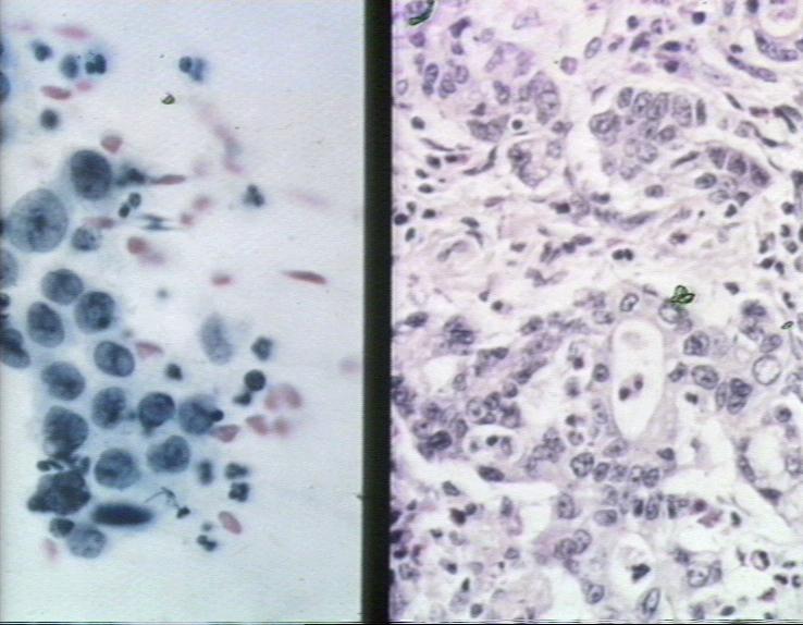
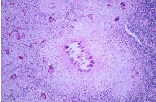
| 11:42, 3 September 2007 | Abnormal mitoses.jpg (file) |  |
17 KB | Abnormal mitoses. Pending permission from Brian Smyth. | 1 |
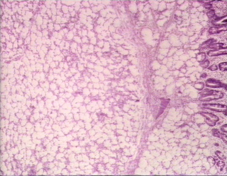
| 18:24, 9 August 2007 | Abomasal lymphoma.jpg (file) |  |
68 KB | 1 | |
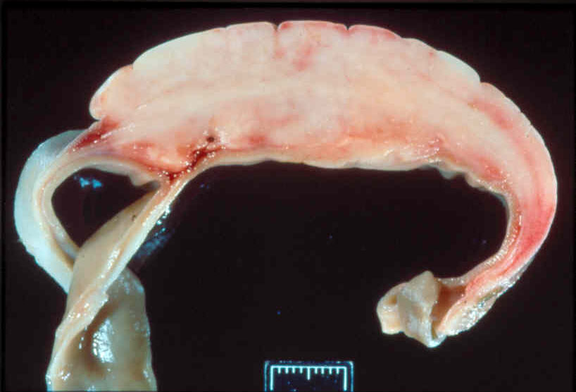
| 21:05, 20 August 2007 | Abscess centre and capsule.jpg (file) |  |
19 KB | Gross appearance of an abscess, showing the necrotic centre and the capsule. Pending permission from Brian Smyth. | 1 |
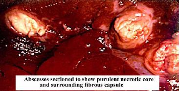
| 21:03, 20 August 2007 | Abscess slice.jpg (file) |  |
31 KB | Slice through and abscess, showing all the layers. Pending permission from Brian Smyth. | 1 |
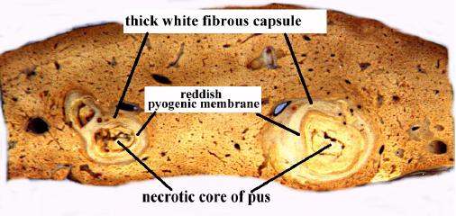
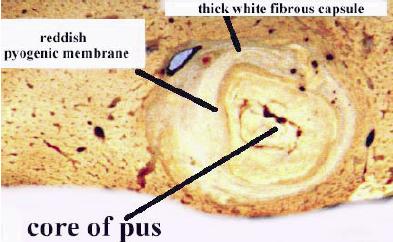
| 09:23, 31 August 2007 | Acute abscess.jpg (file) |  |
23 KB | Cross section through an acute abscess. Pending permission from Brian Smyth. | 1 |
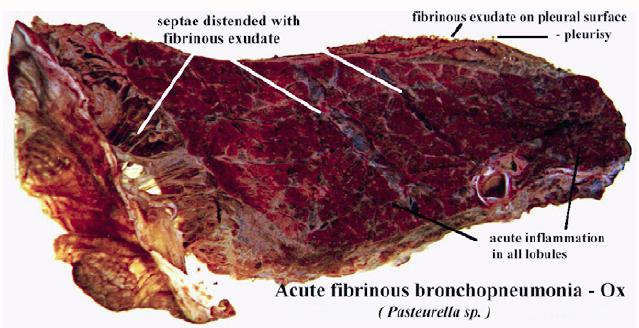
| 09:20, 31 August 2007 | Acute fibrinous bronchopneumonia.jpg (file) |  |
47 KB | Gross appearance on acute fibrinous bronchopneumonia in the ox, caused by ''Pasteurella sp.''. Note the septae are distended with fibrinous exudate. Pending permission from Brian Smyth. | 1 |
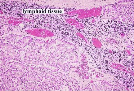
| 11:53, 3 September 2007 | Adenocarcinoma metastasis to lymph node.jpg (file) |  |
52 KB | Histological appearance of lymphatic spread of adenocarcinoma to the lymph nodes. Pending permission from Brian Smyth. | 1 |
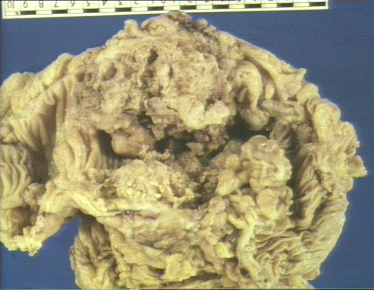
| 18:39, 9 August 2007 | Adenocarcinoma stomach.jpg (file) |  |
69 KB | 1 | |
| 18:39, 9 August 2007 | Adenocarcinoma stomach histopath2.jpg (file) |  |
64 KB | 1 | |
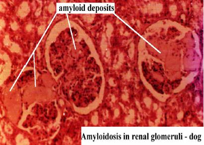
| 19:08, 20 August 2007 | Amyloidosis.jpg (file) |  |
29 KB | Histological appearance of amyloidosis. Pending permission from Brian Smyth. | 1 |
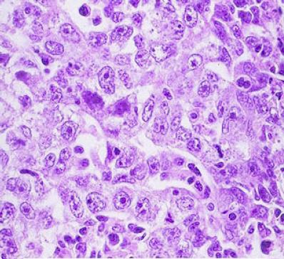
| 12:21, 31 August 2007 | Anaplatic carcinoma.jpg (file) |  |
41 KB | Anaplastic carcinoma. Pending permission from Brian Smyth. | 1 |
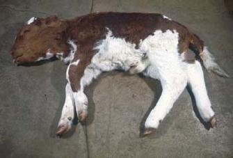
| 16:30, 29 August 2007 | Anasarca.jpg (file) |  |
12 KB | Anasarca. Pending permission from Brian Smyth. | 1 |
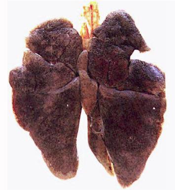
| 12:26, 21 August 2007 | Anthracosis gross.jpg (file) |  |
23 KB | Gross appearance of antrhacosis in the lungs of a dog. Pending permission from Brian Smyth. | 1 |
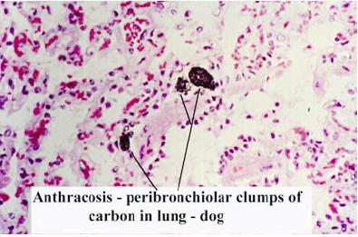
| 12:27, 21 August 2007 | Anthracosis histo.jpg (file) |  |
27 KB | Histological appearance of anthracosis. Pending permission from Brian Smyth. | 1 |
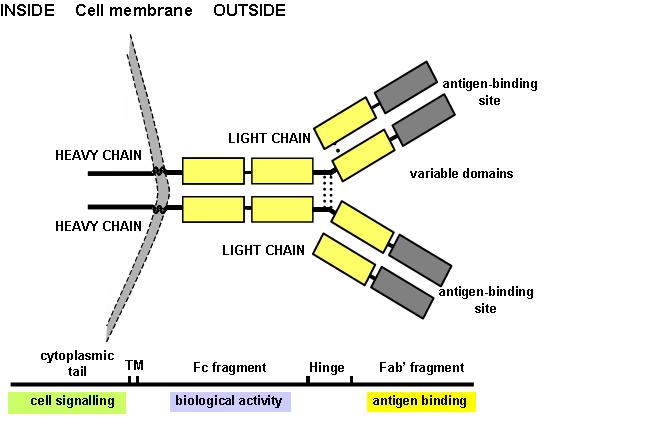
| 16:04, 4 September 2007 | Antibody structure.jpg (file) |  |
34 KB | Diagram showing the basic structure of antibody. Pending permission from John Hopkins. | 1 |
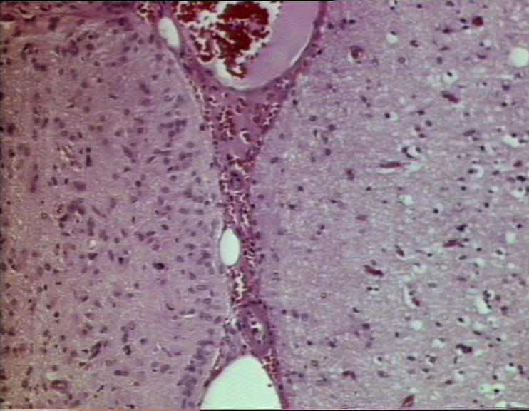
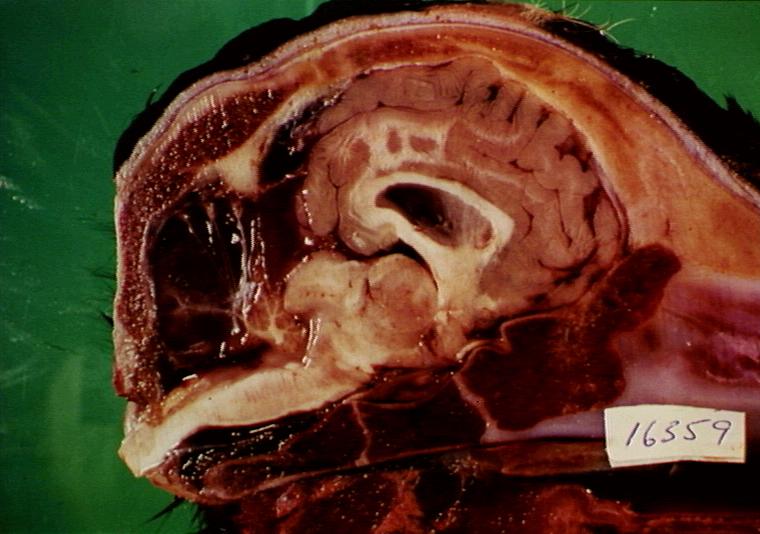
| 18:17, 25 March 2008 | Aqueductstenosis.jpg (file) |  |
74 KB | Aqueduct stenosis, causing hydrocephalus. Courtesy of BioMed Image Archive. | 1 |
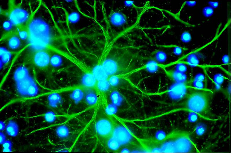
| 16:18, 26 March 2008 | Astrocyte.jpg (file) |  |
75 KB | An astrocyte in culture, stained immunofluorecently. The astrocyte processes are stained green, and the nuclei of this and other cells in the culture are stained blue. Imagae courtesy of [http://www2.unil.ch/edab/old/fr/presse_info.htm the Eurpoean Dana | 1 |
| 11:43, 26 March 2008 | Astrocytomagross.jpg (file) |  |
39 KB | Astrocytoma. Courtesy of BioMed Image Archive | 1 |
| 11:44, 26 March 2008 | Astrocytomahisto.jpg (file) |  |
68 KB | Astrocytoma - histological view. Courtesy of BioMed Image Archive. | 1 |
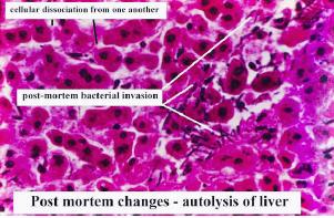
| 10:35, 21 August 2007 | Autolysis of liver.jpg (file) |  |
21 KB | Histological appearance of autolysis in the liver. Pending permission from Brian Smyth. | 1 |
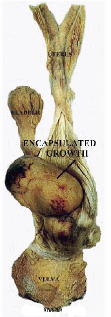
| 11:14, 3 September 2007 | Benign leiomyoma.jpg (file) |  |
25 KB | Benign neoplasm (leiomyoma). Note that the growth is encapsulated. Pending permission from Brian Smyth. | 1 |
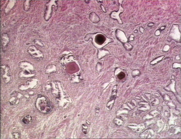
| 12:04, 31 August 2007 | Benign prostatic hyperplasia.jpg (file) |  |
89 KB | Benign prostatic hyperplasia (man). Courtesy of BioMed Archive. | 1 |
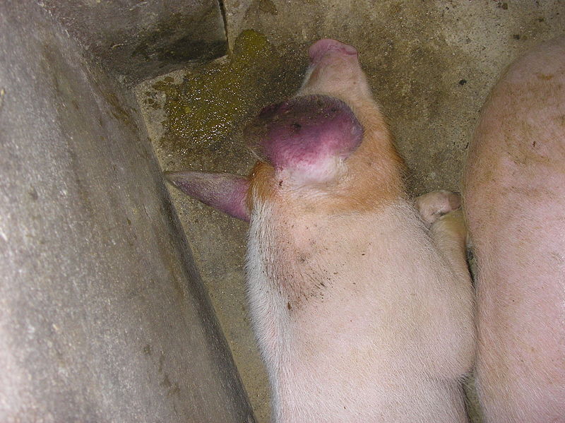
| 20:04, 27 August 2010 | Blue Eared Pig.jpg (file) |  |
112 KB | {{Information |Description=A pig with PRRS showing blue ears. |Source=Wikimedia Commons |Date=Orginally uploaded to Wikimedia Commons 31st December 2007. Uploaded to WikiVet 27th August 2010. |Author=Dingar |Permission=See below }} | 1 |
| 13:03, 20 August 2010 | Bluetongue Virus.gif (file) |  |
272 KB | {{Information |Description= Negatively stained bluetongue virus–like particle that caused a cytopathic effect in BHK-21 cells. Scale bar = 50 nm |Source= Wikimedia Commons |Date=2nd May 2007 |Author=Not Named |Permission=This image is a work of the | 1 |
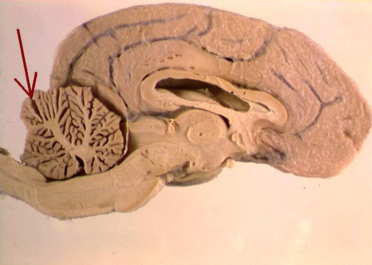
| 14:33, 27 March 2008 | Braincerebellumarrow.jpg (file) |  |
49 KB | Cross section of the brain. The arrow indicates the location of the cerebellum. Courtesy of BioMed Image Archive. | 1 |
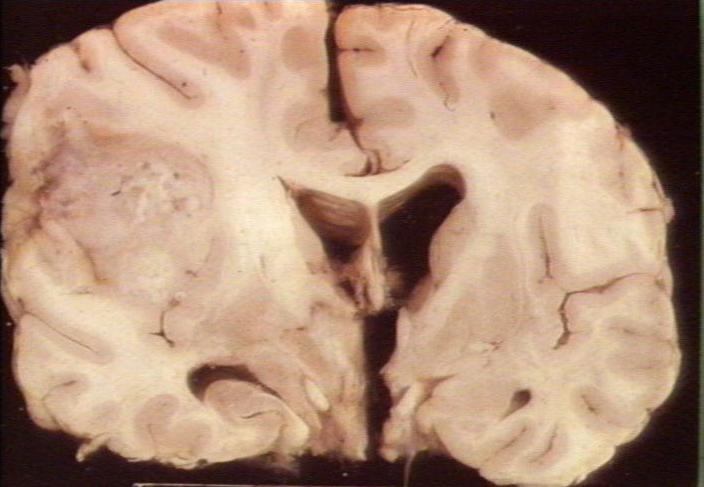
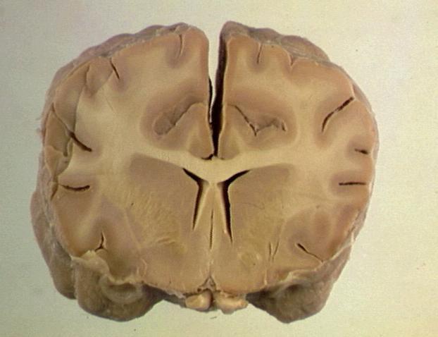
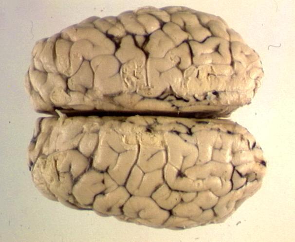
| 13:31, 27 March 2008 | Braincosssection.jpg (file) |  |
31 KB | Cross-section through the brain, showing the cerebrum, basal nuclei and lateral ventricle. The white and grey matter can be easily distinguished. | 1 |
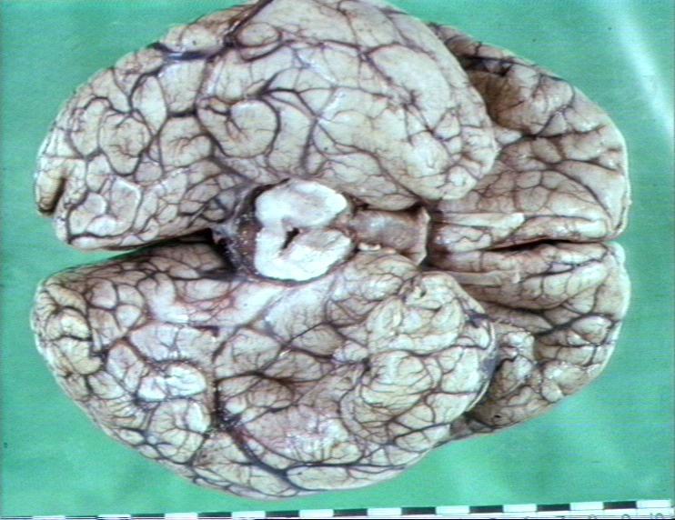
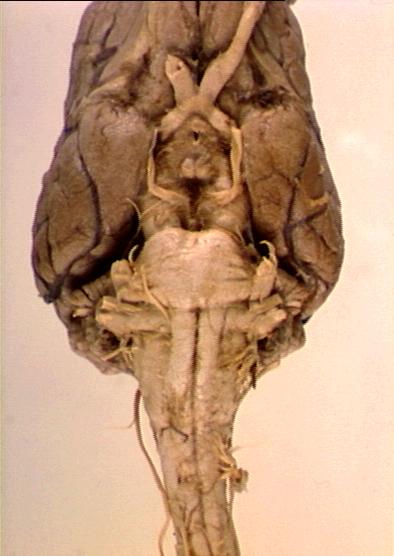
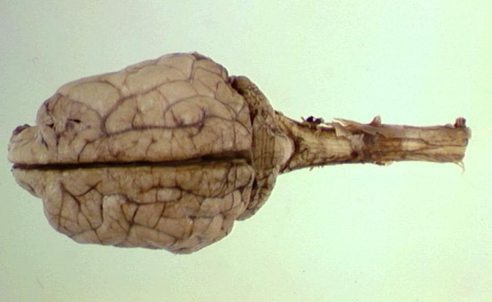
| 12:34, 10 January 2008 | Brainstemcranialnervespyramids.jpg (file) |  |
35 KB | Whole brain (canine) viewed from below showing brain stem, cranial nerves, and pyramids. Courtesy of BioMed Image Archive. | 1 |
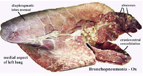
| 10:11, 31 August 2007 | Bronchopneumonia ox.jpg (file) |  |
40 KB | Bronchonpneumonia in the ox (gross). Pending permission from Brian Smyth. | 1 |
| 11:24, 10 August 2007 | Brunner gland adenoma.jpg (file) |  |
75 KB | Adenoma of brunners gland (duodenum). | 1 |
| 21:27, 20 August 2007 | Calcification.jpg (file) |  |
19 KB | Histological appearance of calcification. Pending permission from Brian Smyth. | 1 |
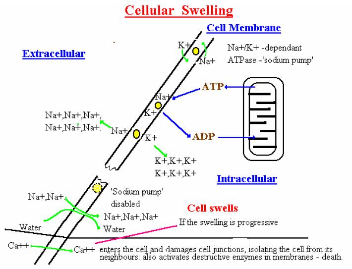
| 18:22, 20 August 2007 | Cellular swelling diagram.jpg (file) |  |
56 KB | The mechanism of cellular swelling. Pending permission from Brian Smyth. | 1 |
| 17:44, 4 January 2008 | Cerebellarhypoplasia.jpg (file) |  |
65 KB | Cross-section of head of seven-day-old calf showing cerebellar hypoplasia and atrophy of occipital cortical lobe. Courtesy of BioMed Image Archive. | 1 |
| 13:28, 27 March 2008 | Cerebralcortex.jpg (file) |  |
39 KB | Whole brain viewed from above showing cerebrum. Courtesy of BioMed Image Archive | 1 |
| 12:26, 10 January 2008 | Cerebrumbrainstemcerebellum.jpg (file) |  |
34 KB | Whole brain (canine) viewed from above showing the cerebrum, brain stem, and cerebellum. Courtesy of BioMed Image Archive | 1 |
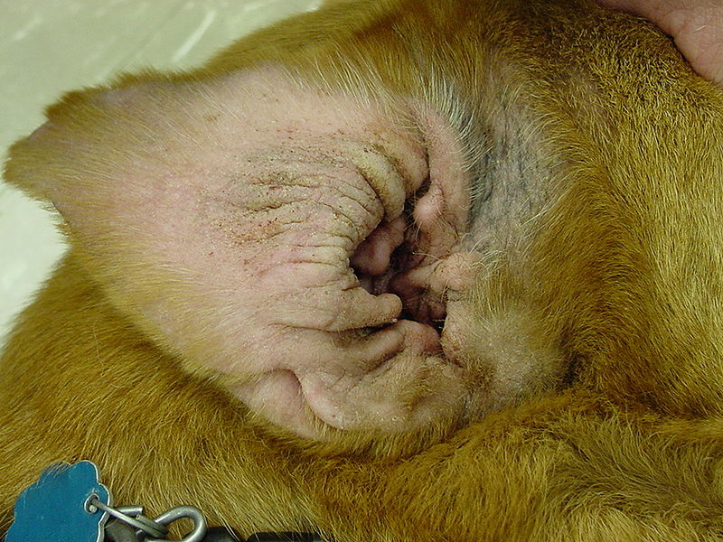
| 19:19, 26 August 2010 | Chronic Allergic Otitis.jpg (file) |  |
140 KB | {{Information |Description=Chronic allergic otitis externa in the dog. |Source=Wikimedia Commons |Date=Originally uploaded to Wikimedia Commons 19th November 2006. Uploaded to WikiVet 26th August 2010. |Author=Caroldermoid |Permission=See below }} | 1 |
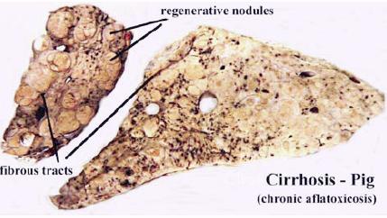
| 10:31, 31 August 2007 | Cirrhosis pig.jpg (file) |  |
25 KB | Cirrhosis, due to chronic liver damage in the pig. Pending permission fro Brian Smyth. | 1 |
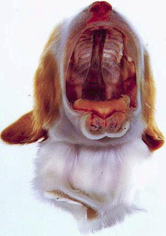
| 15:27, 3 September 2007 | Cleft palate.jpg (file) |  |
21 KB | Cleft palate in the cat. Pending permission from Brian Smyth. | 1 |
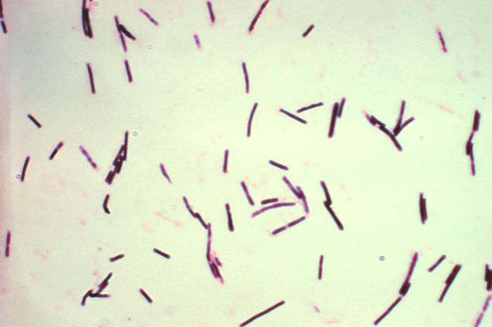
| 19:28, 24 August 2010 | Clostridium perfringens.jpg (file) |  |
28 KB | {{Information |Description=Photomicrograph showing Clostridium perfringens grown in Schaedler’s broth using Gram-stain |Source=Wikimedia Commons |Date=Image created 1974. Originally uploaded to Wikimedia Commons 1st May 2005. Uploaded to WikiVet 24th Au | 1 |
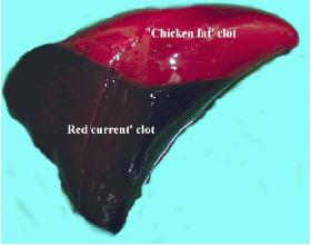
| 09:51, 21 August 2007 | Clot.jpg (file) |  |
8 KB | A post-mortem clot, showing the upper "chicken-fat" clot and the lower "redcurrant-jelly" clot. Pending permission from Brian Smyth. | 1 |
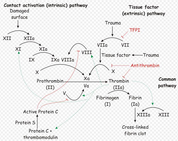
| 15:21, 23 August 2010 | Coagulation Cascade.jpg (file) |  |
89 KB | {{Information |Description= Diagram showinf the coagulation cascade. |Source= Wikimedia Commons |Date=Originally uploaded 22 April 2007 |Author=Joe D |Permission=See below }} | 1 |
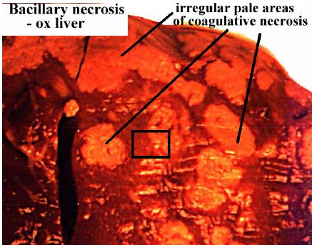
| 20:51, 20 August 2007 | Coagulative necrosis bacillary necrosis.jpg (file) |  |
35 KB | Coagulative necrosis due to bacillary necrosis. Pending permission form Brian Smyth. | 1 |
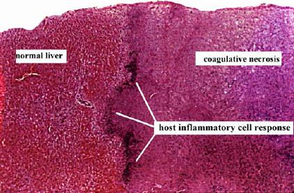
| 20:52, 20 August 2007 | Coagulative necrosis histo.jpg (file) |  |
35 KB | Histological appearance of coagulative necrosis. Pending permission from Brian Smyth. | 1 |
| 09:23, 22 August 2007 | Cocktails.jpg (file) |  |
30 KB | 1 | |
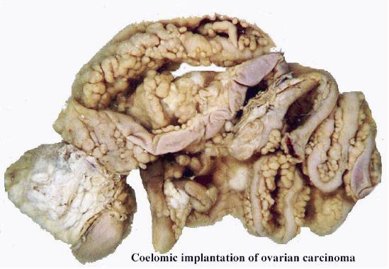
| 11:56, 3 September 2007 | Coelomic implantation ovarian carcinoma.jpg (file) |  |
36 KB | Coelomic implantation of ovarian carcinoma. Pending permission from Brian Smyth. | 1 |
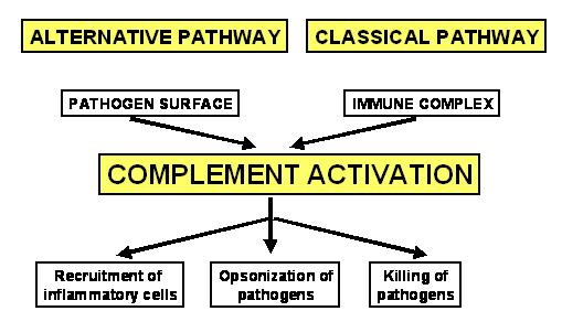
| 10:33, 6 September 2007 | Complement activation.jpg (file) |  |
56 KB | Complement activation. Pending permission from John Hopkins. | 1 |
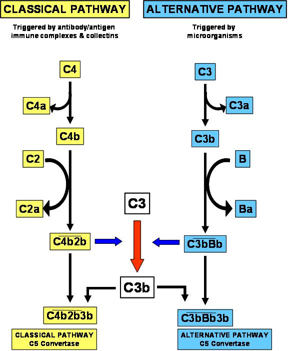
| 10:47, 6 September 2007 | Complement activity.jpg (file) |  |
29 KB | Complement activity. Pending permission from John Hopkins. | 1 |
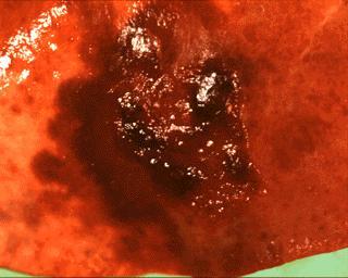
| 10:31, 7 September 2007 | Consolidation and haemorrhage lung.jpg (file) |  |
16 KB | Consolidation and haemorrhage in the lung. Courtesy of BioMed Archive. | 1 |
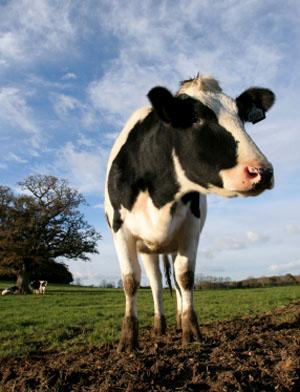
| 16:28, 7 August 2007 | Cow2.jpg (file) |  |
24 KB | 1 | |
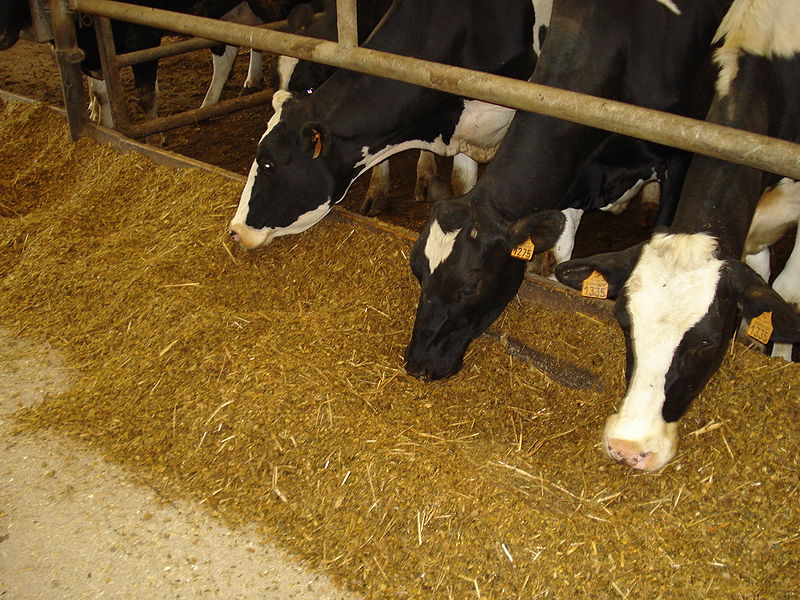
| 07:40, 27 August 2010 | Cows Eating TMR.jpg (file) |  |
144 KB | {{Information |Description=Holstein cattle eating a total mixed ration. |Source=Wikimedia Commons |Date=Originally uploaded to Wikimedia Commons 9th October 2007. Uploaded to WikiVet 27th August 2010. |Author=Tractorboy60 |Permission=See below }} | 1 |
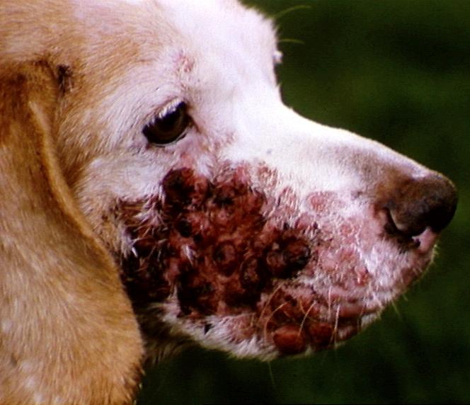
| 10:11, 7 September 2007 | Cutaneous lymphosarcoma.jpg (file) |  |
57 KB | Cutaneous lymphosarcoma. Diffuse lesion on right side of face. Courtesy of BioMed Archive. | 1 |