Uploads by Ctrace
Jump to navigation
Jump to search
This special page shows all uploaded files.
| Date | Name | Thumbnail | Size | Description | Versions |
|---|---|---|---|---|---|
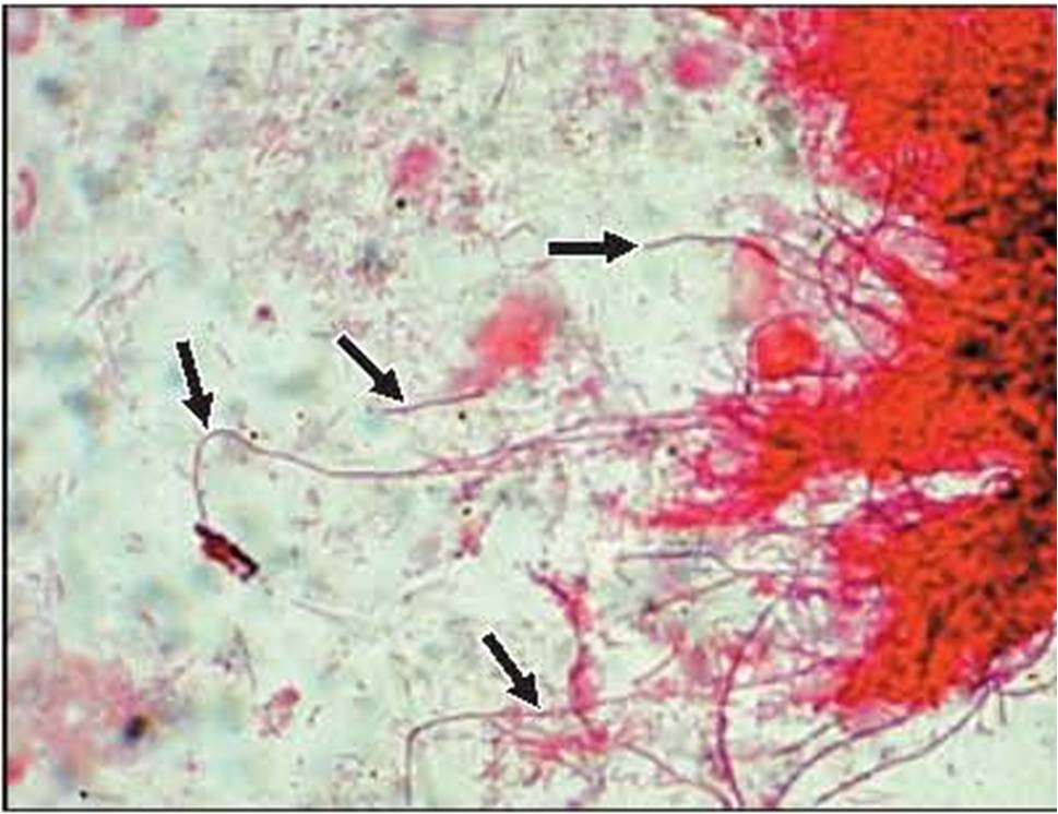
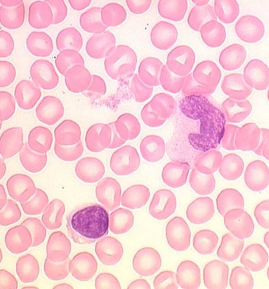
| 12:29, 7 December 2010 | 11.jpg (file) |  |
79 KB | {{Information |Description=cytology Q&A 11 |Source=Mansons |Date=07/12/10 |Author=Mansons |Permission=See below |Other_versions= }} | 1 |
| 13:06, 7 December 2010 | 15.jpg (file) |  |
36 KB | {{Information |Description=test for cytology Q&A |Source=Mansons |Date=07/12/10 |Author=Mansons |Permission=See below |Other_versions= }} | 1 |
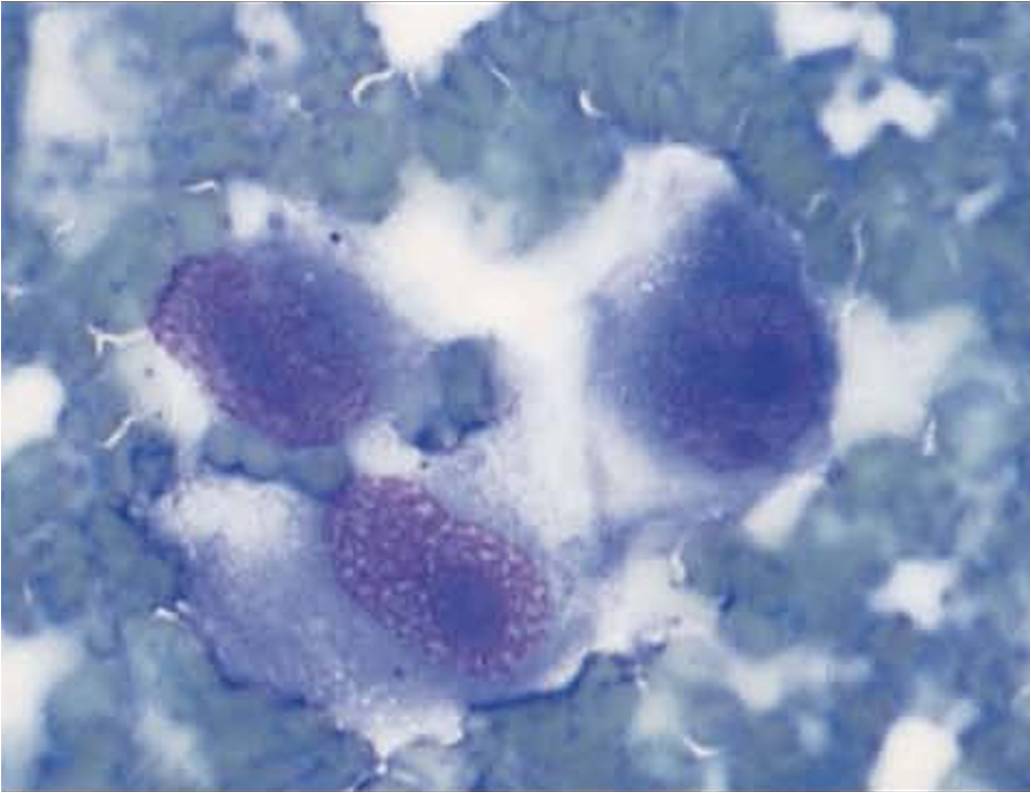
| 11:54, 7 December 2010 | 7.jpg (file) |  |
58 KB | {{Information |Description=test cytology image |Source=Mansons publishing |Date=07/12/10 |Author=Mansons |Permission=See below |Other_versions= }} | 1 |
| 14:41, 31 January 2011 | Addpagetobook.jpg (file) |  |
60 KB | {{Information |Description=Screenshot of 'Add page to book' |Source=WikiVet |Date=31/01/11 |Author=Chris Trace |Permission=See below |Other_versions= }} | 1 |
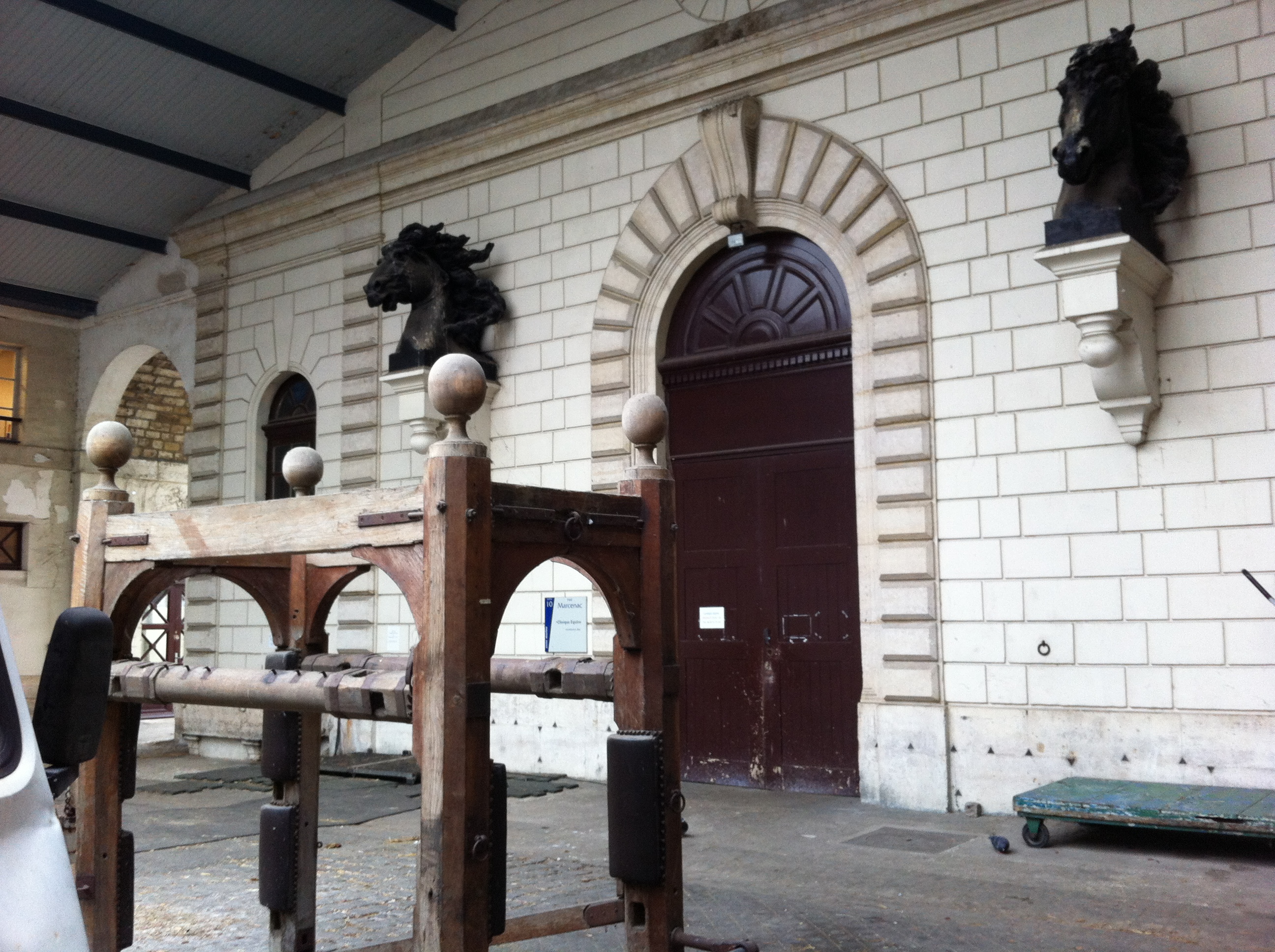
| 17:52, 30 March 2012 | Alfort stables.jpg (file) |  |
1.85 MB | 1 | |
| 13:32, 16 March 2011 | Anatomy video.png (file) |  |
28 KB | {{Information |Description=Logo for the Anatomy section of WikiVideo |Source=WikiVet |Date=16/03/2011 |Author=Chris Trace |Permission=See below |Other_versions= }} | 1 |
| 07:55, 13 April 2011 | April 2001 Issue 4.pdf (file) | 137 KB | {{Information |Description =4th Issue of the WikiVet newsletter |Source =WikiVet |Author =Chris Trace |Date =Sent 7th April 2011 |Permission = |other_versions = }} | 1 | |
| 12:36, 15 November 2011 | Aspark.jpg (file) |  |
7 KB | {{Information |Description ={{en|1=Cropped version of original image}} |Source =File:155003 1630342032382 1051549747 2555529 2021648 n (1).jpg |Author =Alistair Spark |Date =15/11/2011 |Permission = |other_versions = | 1 |
| 12:42, 15 November 2011 | Aspark.png (file) |  |
115 KB | {{Information |Description ={{en|1=cropped version of original image}} |Source =File:155003 1630342032382 1051549747 2555529 2021648 n (1).jpg |Author =Alistair Spark |Date =15/11/2011 |Permission = |other_versions = | 1 |
| 07:55, 30 April 2012 | BSAVAOrange.jpg (file) |  |
39 KB | 1 | |
| 09:28, 24 May 2011 | Blood and Haemopoiesis.swf (file) | 1.9 MB | {{Information |Description =Blood and Haemopoiesis powerpoint tutorial, converted into swf format |Source =Royal Veterinary College |Author =John Bredl |Date =2011 |Permission = | 1 | |
| 14:51, 25 March 2011 | Blood and haemopoiesis.jpg (file) |  |
36 KB | {{Information |Description=Screenshot from Powerpoint, courtesy of the Royal Veterinary College |Source=The Royal Veterinary College |Date=25/03/2011 |Author=Chris Trace |Permission=See below |Other_vers | 1 |
| 11:20, 9 May 2011 | Bone and Cartilage.swf (file) | 1.73 MB | {{Information |Description=PowerPoint tutorial on Bone and Cartilage Histology, converted to swf format. Resource page can be found here |Source=The Royal Veterinary College |Date=2011 |Author=[[RVC|The R | 1 | |
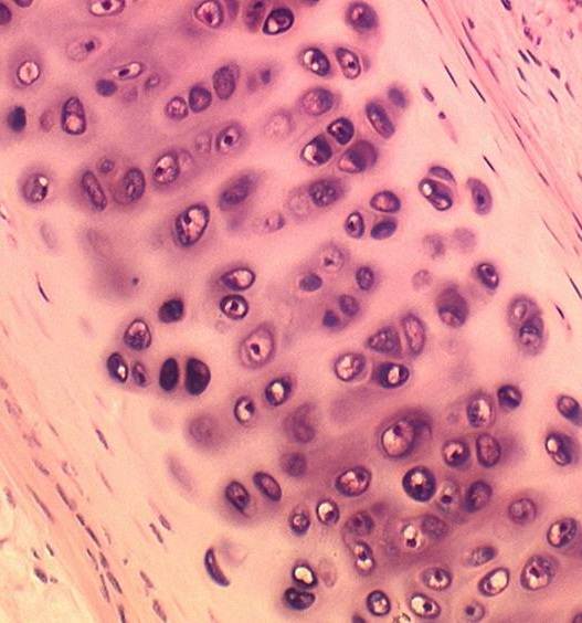
| 11:28, 9 May 2011 | Bone and Cartilage1.jpg (file) |  |
42 KB | {{Information |Description=Screenshot from firstBone and Cartilage Powerpoint tutorial, courtesy of the Royal Veterinary College. Image shows hyaline cartilage from a trachea |Source=[[RVC|The Royal Veter | 1 |
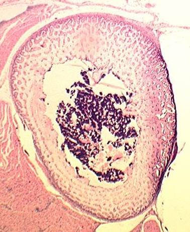
| 12:26, 9 May 2011 | Bone and Cartilage2.jpg (file) |  |
38 KB | {{Information |Description=Screenshot from secondBone and Cartilage Powerpoint tutorial, courtesy of the Royal Veterinary College. Image shows transverse section of diaphysis of developing rabbit bone |So | 1 |
| 12:19, 9 May 2011 | Bone and Cartilage2.swf (file) | 1.88 MB | {{Information |Description=2nd PowerPoint tutorial on Bone and Cartilage Histology, converted to swf format. Resource page can be found here |Source=The Royal Veterinary College |Date=2011 |Author=[[RVC|T | 1 | |
| 14:28, 8 February 2011 | Book.gif (file) |  |
4 KB | {{Information |Description=Image for eBooks on WikiVet |Source=RVC |Date=08/02/11 |Author=Chris Trace |Permission=See below |Other_versions= }} | 1 |
| 09:58, 23 February 2011 | BovinePregnantUterus.png (file) |  |
1.23 MB | {{Information |Description=screenshot of a Bovine Anatomy video podcast |Source=http://stream2.rvc.ac.uk/Anatomy/bovine/cow-uterus2.wmv |Date=23/02/2011 |Author=Chris Trace |Permission=See below |Other_versions= }} | 1 |
| 13:14, 16 March 2011 | Bovine video.png (file) |  |
26 KB | {{Information |Description=Logo for Bovine video section of WikiVideo |Source=WikiVet |Date=16/03/2011 |Author=Chris Trace |Permission=See below |Other_versions= }} | 1 |
| 12:29, 31 May 2012 | Bovril.png (file) |  |
541 KB | 1 | |
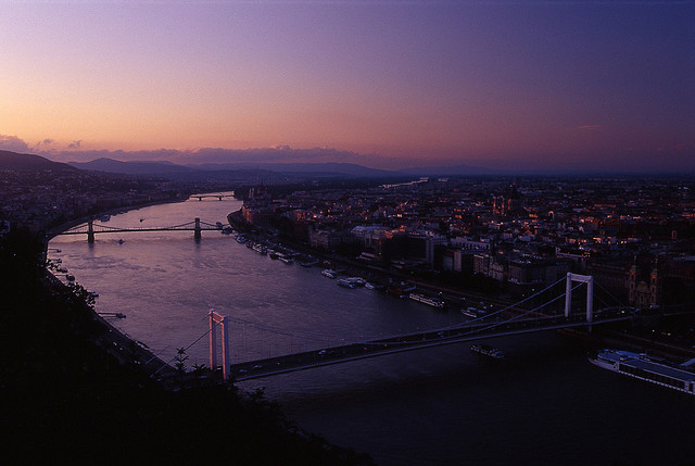
| 13:18, 4 May 2011 | Budapest.jpg (file) |  |
65 KB | {{Information |Description =Photo of Budapest |Source =[http://www.flickr.com/photos/9702937@N06/1418473231/ Flickr] |Author =mck...'s |Date =4th August 2007 |Permission = |other_versions = }} Licensed under a [http://crea | 1 |
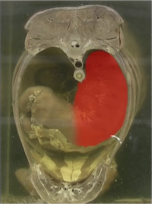
| 09:46, 23 February 2011 | CanineAbdomen.png (file) |  |
755 KB | {{Information |Description=A screenshot of a potcast of the canine abdomen |Source=http://stream2.rvc.ac.uk/Anatomy/canine/Pot0036.wmv |Date=23/02/2011 |Author=Chris Trace |Permission=See below |Other_versions= }} | 1 |
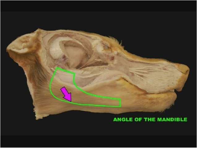
| 10:41, 23 February 2011 | CanineHead.png (file) |  |
305 KB | {{Information |Description=A screenshot of a Canine lateral head potcast |Source=http://stream2.rvc.ac.uk/Anatomy/canine/head_neck/Pot0220.wmv |Date=23/02/2011 |Author=Chris Trace |Permission=See below |Other_versions= }} | 1 |
| 13:19, 16 March 2011 | Canine video.png (file) |  |
27 KB | {{Information |Description=logo for Canine section of WikiVideo |Source=WikiVet |Date=16/03/2011 |Author=Chris Trace |Permission=See below |Other_versions= }} | 1 |
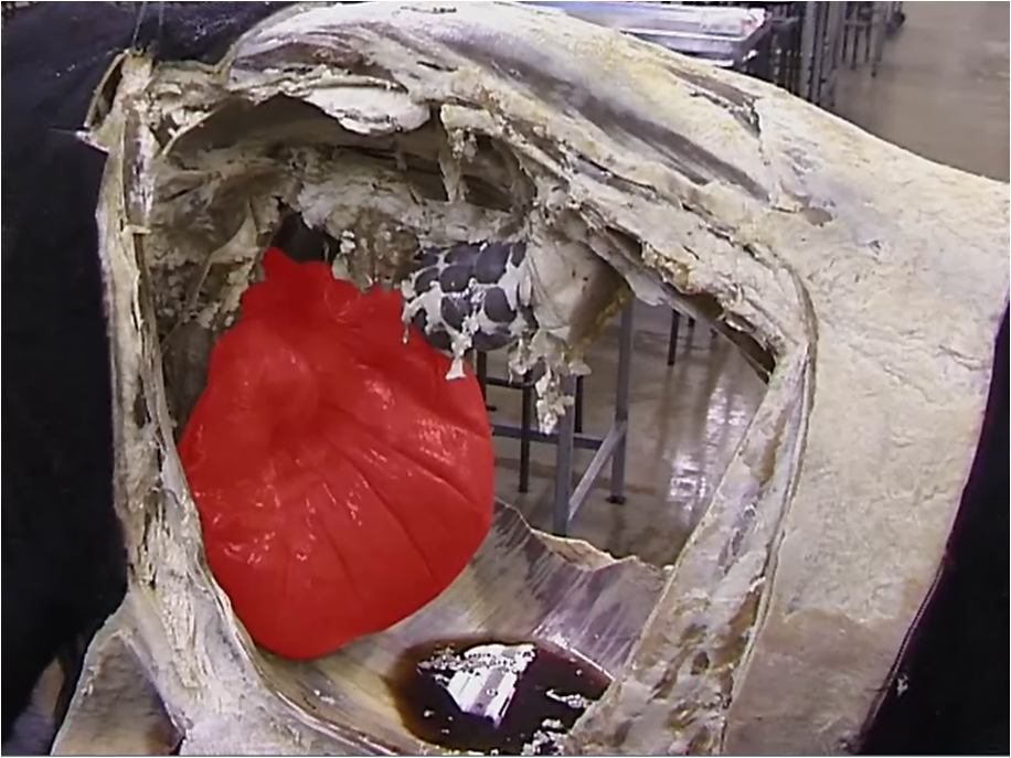
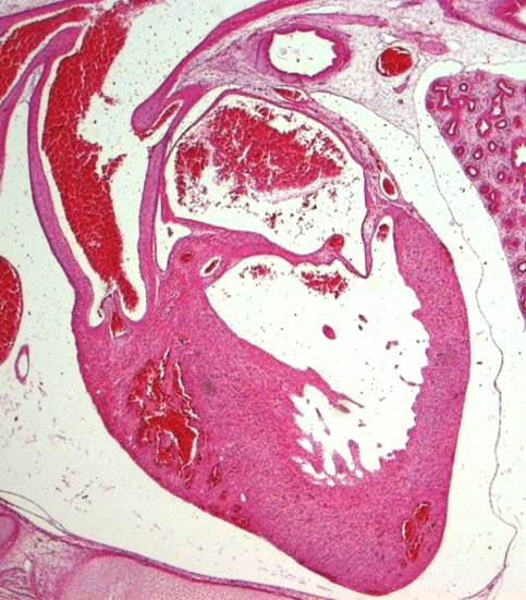
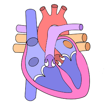
| 16:48, 8 May 2011 | Cardiovascular System.jpg (file) |  |
57 KB | {{Information |Description=Screenshot from Powerpoint, courtesy of the Royal Veterinary College. Shows a longitudinal histological section of a heart |Source=The Royal Veterinary College |Date= | 1 |
| 12:38, 26 June 2011 | Cardiovascular logo.png (file) | 42 KB | Cropped version for use in frontpage templates | 2 | |
| 16:41, 8 May 2011 | Cardiovascular system.swf (file) | 2 MB | {{Information |Description=PowerPoint tutorial on cardiovascular system histology, converted to swf format. Resource page can be found here |Source=The Royal Veterinary College |Date=2011 |Author=[[RVC | 1 | |
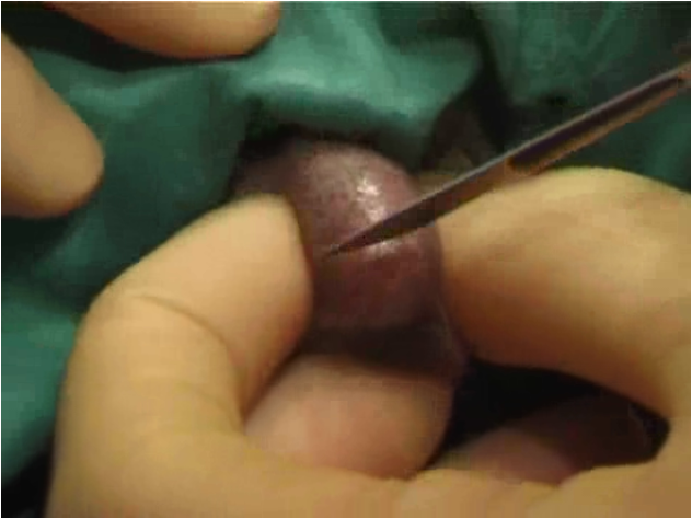
| 10:40, 21 February 2011 | CatCastrate.png (file) |  |
459 KB | {{Information |Description=screenshot of cat castrate video |Source=http://stream2.rvc.ac.uk/Surgical_Skills/cat/castrate/Cat_Castrate_long.wmv |Date=21/02/2011 |Author=Chris Trace |Permission=See below |Other_versions= }} | 1 |
| 11:34, 16 March 2011 | CatSpay.png (file) |  |
539 KB | {{Information |Description=screenshot from Cat Spay video |Source=The Royal Veterinary College |Date=16/03/2011 |Author=Chris Trace |Permission=See below |Other_versions= }} | 1 |
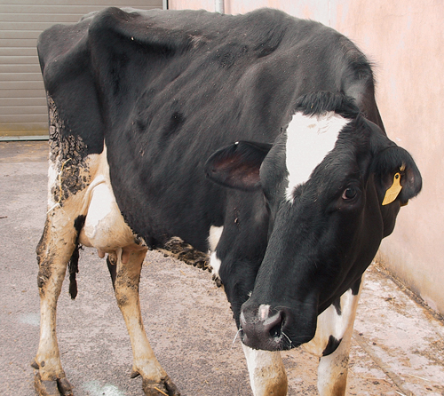
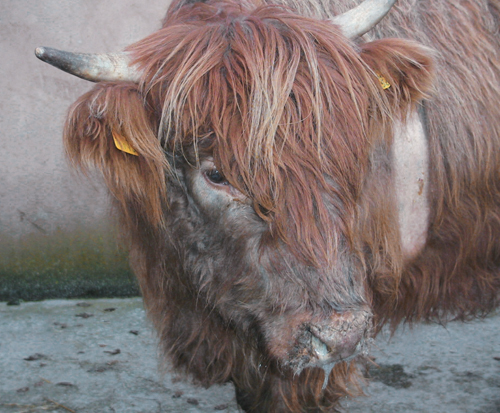
| 17:33, 1 April 2011 | Cattle Medicine 1.jpg (file) |  |
259 KB | {{Information |Description=Image from [http://www.mansonpublishing.com/vet_titles/ScottSACR.html 'Cattle and Sheep Medicine'], with permission from Manson Publishing, as part of the OVAL Project. This is an image of a Ketotic cow |Source=[ | 1 |
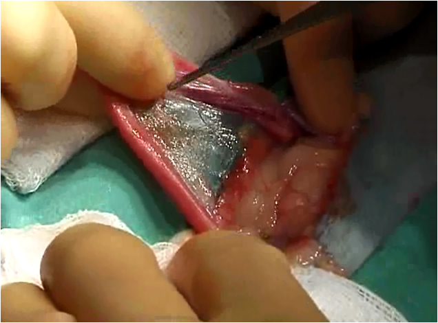
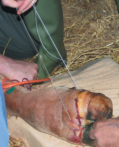
| 17:52, 1 April 2011 | Cattle Medicine 2.jpg (file) |  |
332 KB | {{Information |Description=Image from [http://www.mansonpublishing.com/vet_titles/ScottSACR.html 'Cattle and Sheep Medicine'], with permission from Manson Publishing, as part of the OVAL Project. This is an image of a digit being amputated | 1 |
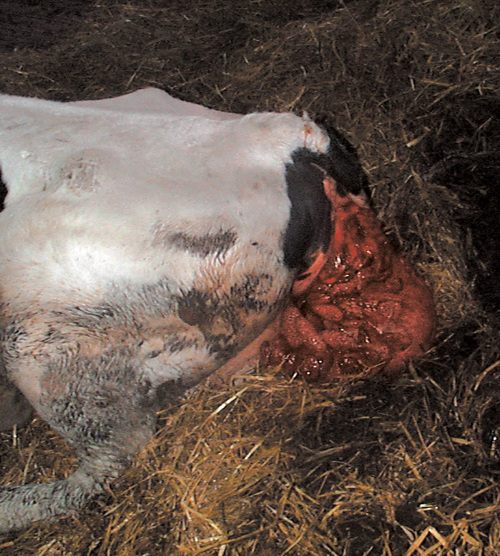
| 18:07, 1 April 2011 | Cattle Medicine 3.jpg (file) |  |
356 KB | {{Information |Description=Image from [http://www.mansonpublishing.com/vet_titles/ScottSACR.html 'Cattle and Sheep Medicine'], with permission from Manson Publishing, as part of the OVAL Project. This is an image of a Uterine Prolapse |Sou | 1 |
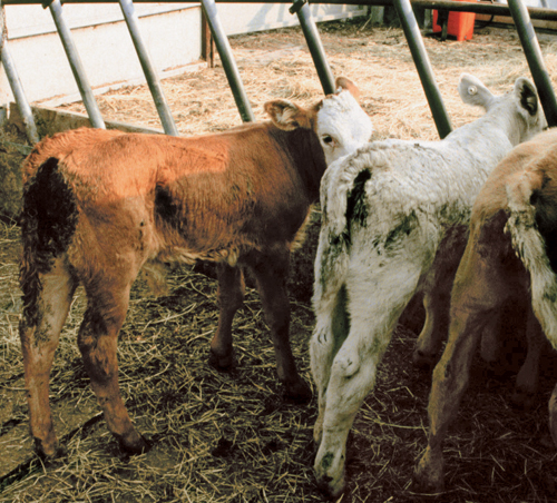
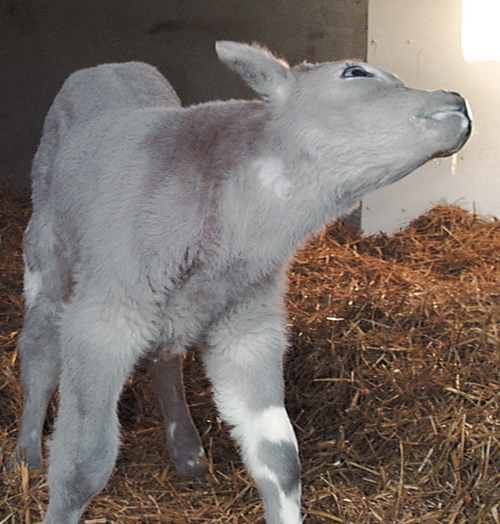
| 18:20, 1 April 2011 | Cattle Medicine 4.jpg (file) |  |
315 KB | {{Information |Description=Image from [http://www.mansonpublishing.com/vet_titles/ScottSACR.html 'Cattle and Sheep Medicine'], with permission from Manson Publishing, as part of the OVAL Project. This is an image of a group of calves with | 1 |
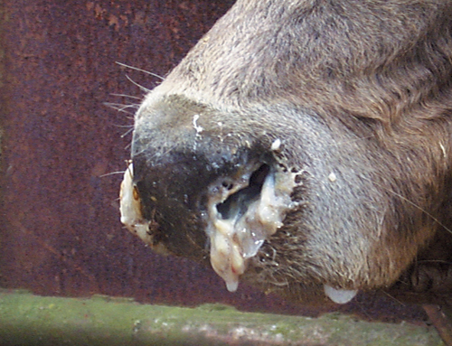
| 18:34, 1 April 2011 | Cattle Medicine 5.jpg (file) |  |
231 KB | {{Information |Description=Image from [http://www.mansonpublishing.com/vet_titles/ScottSACR.html 'Cattle and Sheep Medicine'], with permission from Manson Publishing, as part of the OVAL Project. This is an image of a cow with bilateral na | 1 |
| 18:44, 1 April 2011 | Cattle Medicine 6.jpg (file) |  |
225 KB | {{Information |Description=Image from [http://www.mansonpublishing.com/vet_titles/ScottSACR.html 'Cattle and Sheep Medicine'], with permission from Manson Publishing, as part of the OVAL Project. This is an image of a cow with nasal discha | 1 |
| 15:01, 5 April 2011 | Cattle Medicine 7.jpg (file) |  |
282 KB | {{Information |Description =Image from [http://www.mansonpublishing.com/vet_titles/ScottSACR.html 'Cattle and Sheep Medicine'], with permission from Manson Publishing, as part of the OVAL Project. This is an image of a calf with Meningi | 1 |
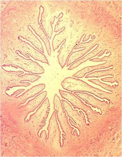
| 11:18, 6 June 2011 | Cervix.jpg (file) |  |
43 KB | {{Information |Description=Screenshot from Powerpoint, courtesy of the Royal Veterinary College. Shows transverse histological section through cervix. |Source=The Royal Veterinary College | | 1 |
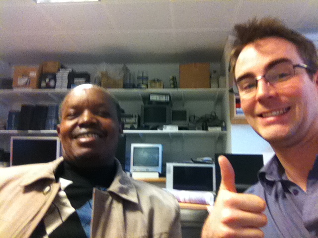
| 16:59, 18 April 2012 | Chris and Paul.jpg (file) |  |
132 KB | 1 | |
| 11:56, 10 March 2012 | ChrisandMarmite.jpg (file) |  |
24 KB | 1 | |
| 11:34, 19 May 2011 | ComparativeLivers.jpg (file) |  |
37 KB | {{Information |Description =Image of a ruminant, equine, canine and porcine liver. |Source =Murcia Vet School |Author =[[Spain - Universidad de Murcia Facultad de Veterinaria|Mur | 1 |
| 11:23, 19 May 2011 | Comparative stomachs.jpg (file) |  |
42 KB | {{Information |Description =Image of a ruminant abomasum and an equine, canine and porcine stomach. |Source =Murcia Vet School |Author =[[Spain - Universidad de Murcia Facultad d | 1 |
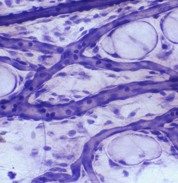
| 17:39, 18 March 2011 | Connective Tissue.png (file) |  |
1.01 MB | {{Information |Description=Screenshot from Powerpoint, courtesy of the Royal Veterinary College |Source=The Royal Veterinary College |Date=18/03/2011 |Author=Chris Trace |Permission=See below |Other | 1 |
| 08:49, 24 May 2011 | Connective Tissue.swf (file) | 1.99 MB | {{Information |Description =PowerPoint tutorial on Connective Tissue Histology, converted to swf format. Resource page can be found here |Source =The Royal Veterinary College |Author =Jo | 1 | |
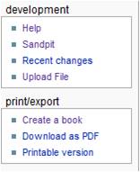
| 14:20, 31 January 2011 | Createabook.jpg (file) |  |
6 KB | {{Information |Description=screenshot of 'create a book' function |Source=wikivet.net |Date=31/01/11 |Author=Chris Trace |Permission=See below |Other_versions= }} | 1 |
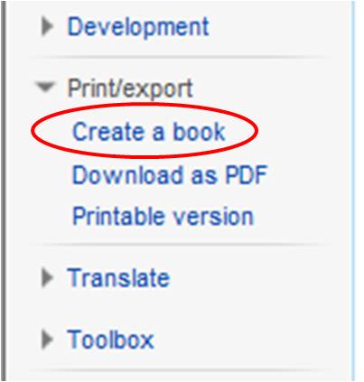
| 14:59, 19 December 2011 | Createabook2.jpg (file) |  |
16 KB | Updated image showing user's skin | 2 |
| 10:40, 1 March 2011 | David Hogg.jpg (file) |  |
20 KB | {{Information |Description=Headshot of David Hogg |Source=RVC |Date=01/03/2011 |Author=Chris Trace |Permission=See below |Other_versions= }} | 1 |
| 15:20, 7 December 2010 | Digital slide box.jpg (file) |  |
282 KB | {{Information |Description=screenshot of the digital slidebox |Source=www.rvc.ac.uk/review |Date=07/12/10 |Author=RVC |Permission=See below |Other_versions= }} | 1 |
| 12:57, 29 October 2013 | Discipline.png (file) |  |
31 KB | 1 | |
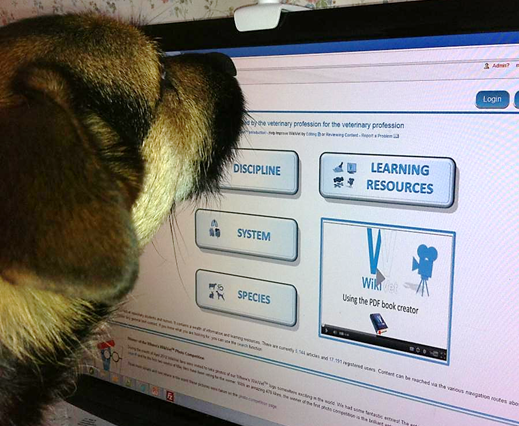
| 16:14, 30 March 2012 | Dogandwikivet.jpg (file) |  |
57 KB | 1 | |
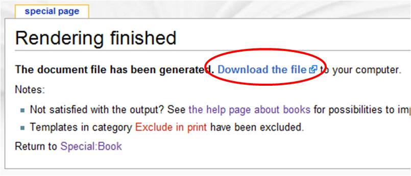
| 15:31, 31 January 2011 | Downloadfile.jpg (file) |  |
31 KB | {{Information |Description=Screenshot of the 'download file' screen after creating a book |Source=WikiVet |Date=31/01/11 |Author=Chris Trace |Permission=See below |Other_versions= }} | 1 |
.jpg)