Uploads by Bara
Jump to navigation
Jump to search
This special page shows all uploaded files.
| Date | Name | Thumbnail | Size | Description | Versions |
|---|---|---|---|---|---|
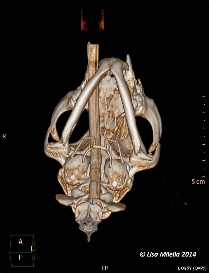
| 15:59, 10 September 2014 | Zygomatic arch fracture.jpg (file) |  |
70 KB | {{Information |Description ={{en|1=Zygomatic arch fracture}} |Source =Lisa Milella |Author =Lisa Milella |Date =2014 |Permission = |other_versions = }} | 1 |
| 15:11, 3 February 2011 | Zoe Belshaw team.jpg (file) |  |
65 KB | {{Information |Description=Zoe Belshaw |Source=own |Date=2011 |Author=Z. Belshaw |Permission=See below |Other_versions= }} | 1 |
| 18:22, 25 January 2011 | Zoe Belshaw.jpg (file) |  |
164 KB | {{Information |Description=Zoe Belshaw |Source=Zoe Belshaw |Date=2011 |Author=Zoe Belshaw |Permission=See below |Other_versions= }} | 1 |
| 18:43, 23 December 2013 | Xmas site.jpg (file) |  |
52 KB | {{Information |Description ={{en|1=Christmas message}} |Source =WikiVet Team |Author =WikiVet Team |Date =December 2013 |Permission = |other_versions = }} | 1 |
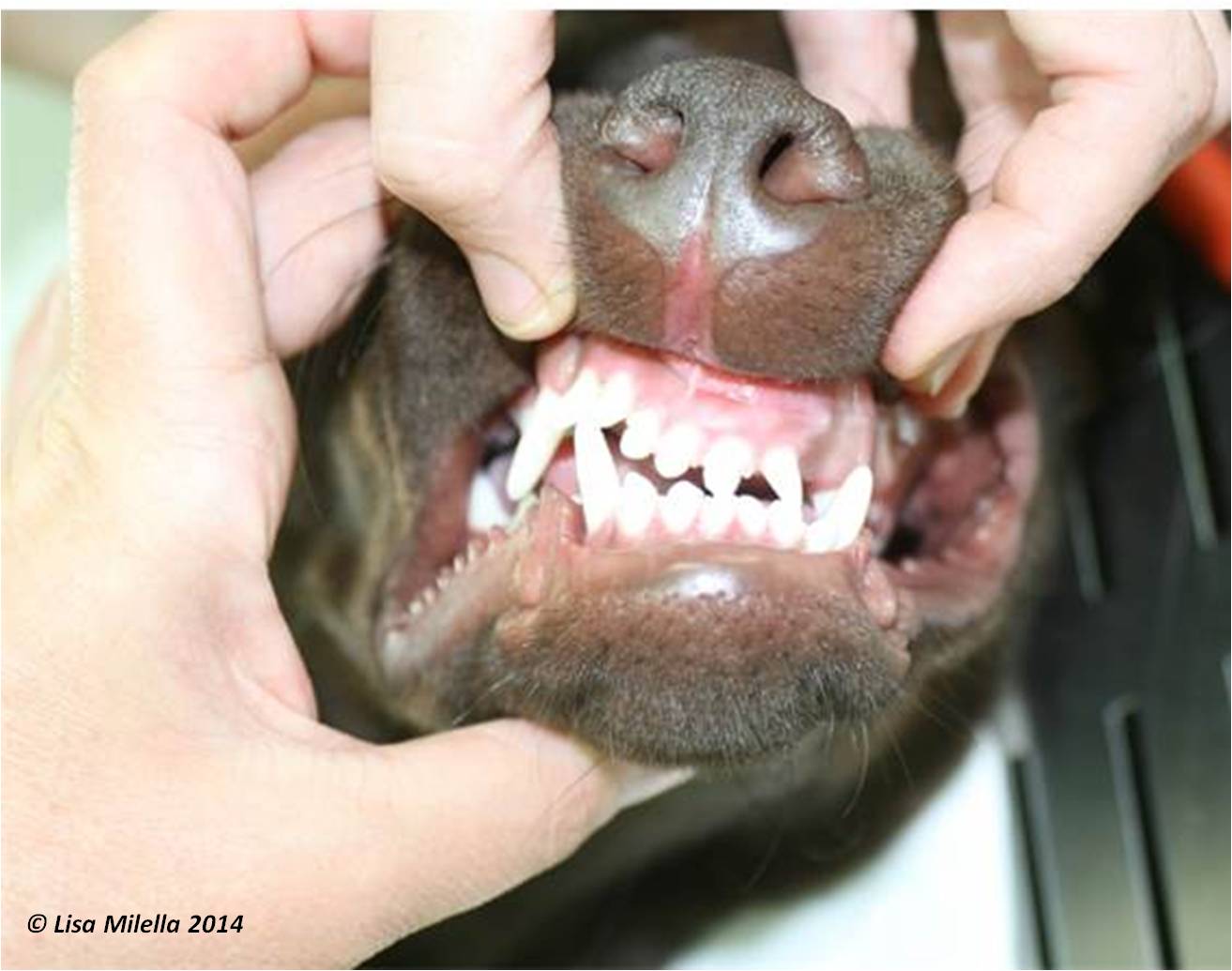
| 15:13, 13 August 2014 | Wry bite 2.jpg (file) |  |
91 KB | better version | 1 |
| 15:12, 13 August 2014 | Wry bite 1.jpg (file) |  |
63 KB | better version | 1 |
| 18:20, 3 January 2010 | Wounds table.jpg (file) |  |
46 KB | Courtesy of The Donkey Sanctuary | 1 |
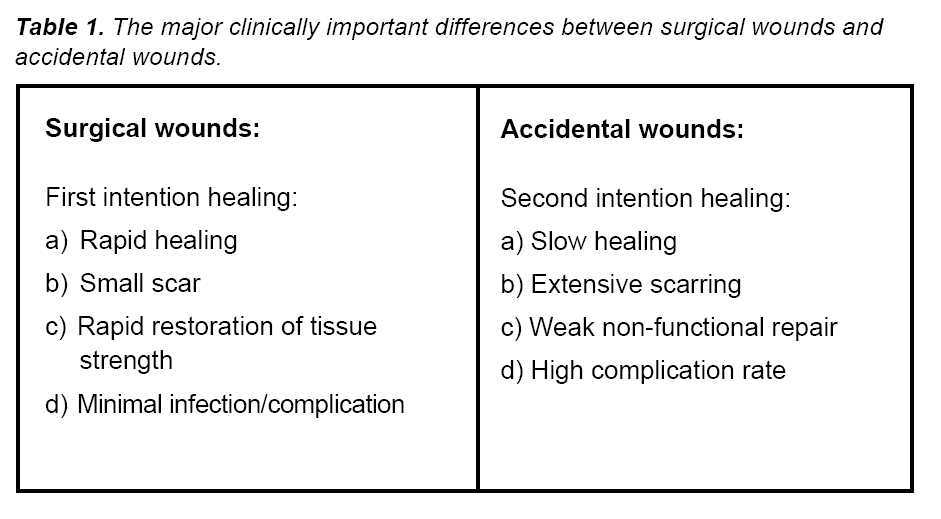
| 18:23, 3 January 2010 | Wound sarcoid.jpg (file) |  |
7 KB | Courtesy of The Donkey Sanctuary | 1 |
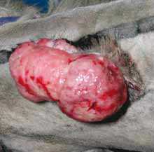
| 18:19, 3 January 2010 | Wound healing.jpg (file) |  |
5 KB | Courtesy of The Donkey Sanctuary | 1 |
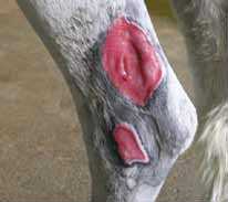
| 18:19, 3 January 2010 | Wound debridement.jpg (file) |  |
7 KB | Courtesy of The Donkey Sanctuary | 1 |
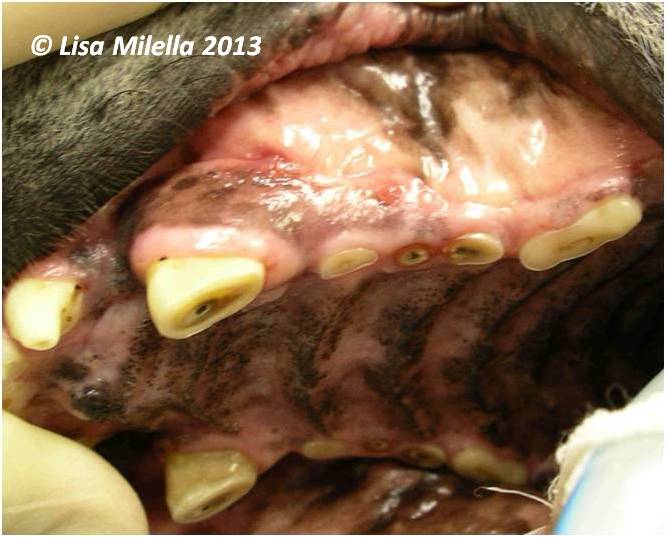
| 13:40, 6 August 2013 | Worn dog teeth.jpg (file) |  |
47 KB | {{Information |Description ={{en|1=Severely worn maxillary canine and premolars. The canine and 2nd premolar have pulp exposure (black spot) and draining fistulae just above the mucogingival junction. The first and 3rd premolars are worn with tertiary | 1 |
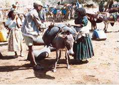
| 22:42, 9 December 2009 | Working donkeys.jpg (file) |  |
10 KB | Courtesy of The Donkey Sanctuary | 1 |
| 18:21, 23 April 2012 | With-search.png (file) | 36 KB | {{Information |Description ={{en|1=potential navigation bar}} |Source =Bara |Author =Bara |Date =April 2012 |Permission = |other_versions = }} | 1 | |
| 08:01, 6 May 2015 | Wiley Logo.jpg (file) | 568 KB | testing | 3 | |
| 12:50, 12 August 2011 | Wikivet hints and tips from Steph.pdf (file) | 50 KB | {{Information |Description ={{en|1=Thoughts on working for WikiVet and the OVAL project.}} |Source =Stephanie Massey |Author =Stephanie Massey |Date =July 2011 |Permission = |other_versions = }} | 1 | |
| 15:19, 1 July 2013 | Wikivet Factoid 1.png (file) |  |
11 KB | {{Information |Description ={{en|1=Image for factoid pages}} |Source =WikiVet |Author =Bara |Date =July 2013 |Permission = |other_versions = }} | 1 |
| 12:13, 8 July 2011 | Wikimedia Commons Image Use on WikiVet.pdf (file) | 884 KB | {{Information |Description ={{en|1=Instruction for uploading an image from Wikimedia Commons}} |Source =Own work |Author =Bara |Date =July 2011 |Permission = |other_versions = }} | 1 | |
| 19:58, 9 November 2012 | WikiQuiz-vetlogic.png (file) |  |
60 KB | {{Information |Description ={{en|1=Logo for Vetlogic sample quizzes}} |Source =WikiVet and Vetlogic |Author =Bara |Date =November 2012 |Permission = |other_versions = }} | 1 |
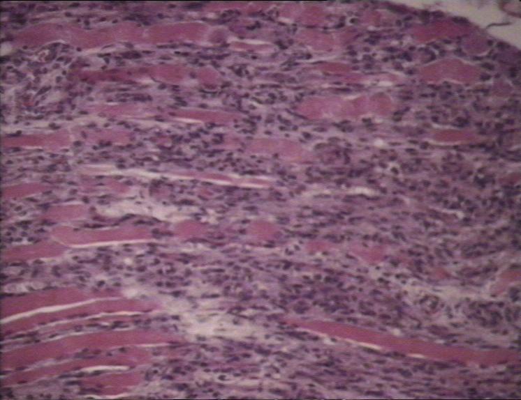
| 12:28, 9 October 2007 | White muscle disease histo.jpg (file) |  |
64 KB | 1 | |
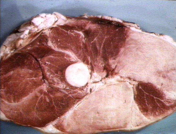
| 12:40, 9 October 2007 | White muscle disease.jpg (file) |  |
66 KB | 1 | |
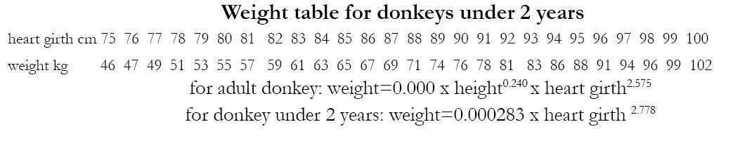
| 22:34, 12 February 2010 | Weight table donkey.jpg (file) | 29 KB | Courtesy of The Donkey Sanctuary | 1 | |
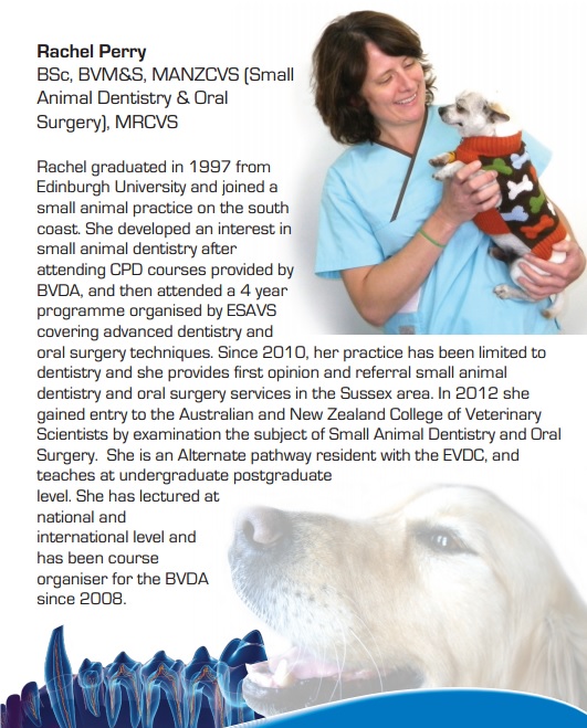
| 16:16, 14 October 2014 | Webinar Rachel Perry.jpg (file) |  |
154 KB | {{Information |Description ={{en|1=Rachel Perry dentistry webinars}} |Source =Mars Petcare |Author =Mars Petcare |Date =2013 |Permission = |other_versions = }} | 1 |
| 15:15, 21 October 2014 | Waltham logo.jpg (file) | 141 KB | {{Information |Description ={{en|1=Waltham logo}} |Source =Mars Petcare |Author =Waltham |Date =2014 |Permission = |other_versions = }} | 1 | |
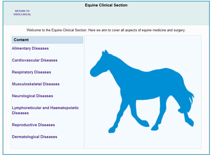
| 09:46, 18 March 2011 | WV navigation.png (file) |  |
55 KB | {{Information |Description=Screen shot of a navigation page |Source=WikiVet |Date=March 2011 |Author=B. Stanikova |Permission=See below |Other_versions= }} | 1 |
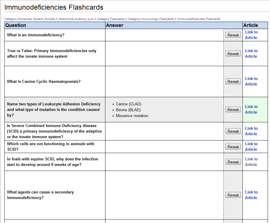
| 09:50, 18 March 2011 | WV flashcard.png (file) |  |
54 KB | {{Information |Description=WikiVet flashcard screen shot |Source=WikiVet |Date=March 2011 |Author=B. Stanikova |Permission=See below |Other_versions= }} | 1 |
| 15:27, 18 January 2012 | WV Path Facebook logo.jpg (file) | 16 KB | {{Information |Description ={{en|1=logo for WikiVet Pathology on Facebook}} |Source =Chris Trace |Author =Chris Trace |Date =January 2012 |Permission = |other_versions = }} | 1 | |
| 14:58, 31 January 2012 | WV Pages.png (file) |  |
32 KB | Changed "articles" to "encyclopaedia" | 4 |
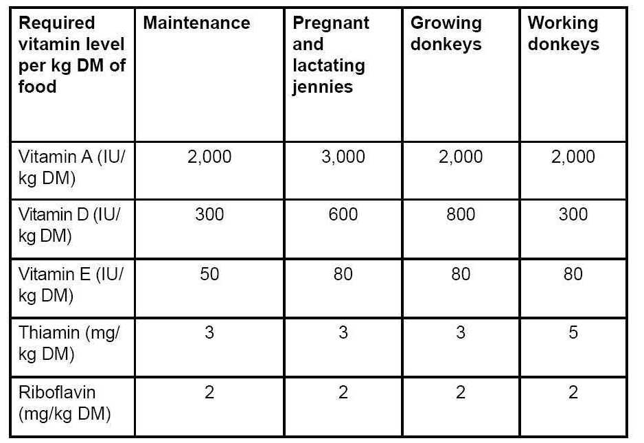
| 18:51, 13 November 2009 | Vitamin table donkeys.jpg (file) |  |
60 KB | Courtesy of The Donkey Sanctuary | 1 |
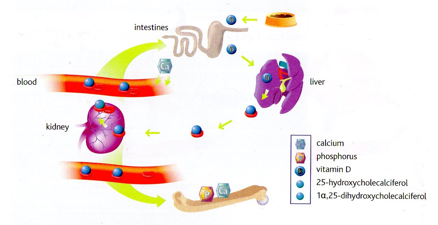
| 09:50, 21 April 2015 | Vitamin D.jpg (file) |  |
229 KB | {{Information |Description ={{en|1=Role of vitamin D in the body. To be active in the body, it must be modified, first in the liver, then in the kidney.}} |Source =Waltham |Author =Waltham |Date =April 2015 |Permission = | | 1 |
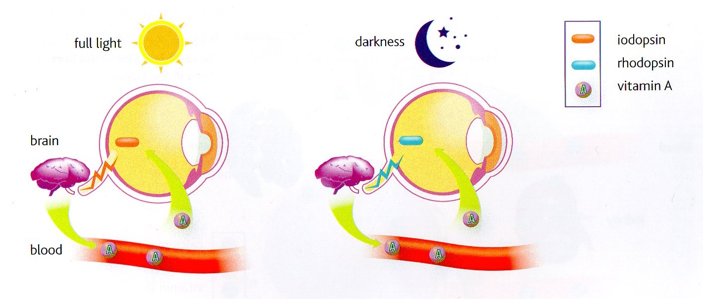
| 09:44, 21 April 2015 | Vitamin A.jpg (file) |  |
199 KB | {{Information |Description ={{en|1=Vitamin A (retinol) plays a role in the synthesis of the retinal pigments needed for the perception of colour (iodopsin of the cones) and night vision (rhodopsin of the rods).}} |Source =Waltham |Author | 1 |
| 12:49, 27 April 2012 | Virology.png (file) |  |
25 KB | {{Information |Description ={{en|1=Virology button}} |Source =Bara |Author =Bara |Date =April 2012 |Permission = |other_versions = }} | 1 |
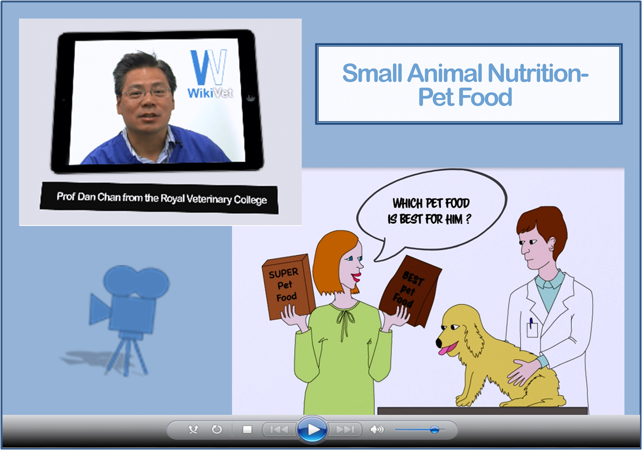
| 15:12, 10 May 2017 | Video screenshots mock up.png (file) |  |
851 KB | Collage of screenshots from the introductory video to Small Animal Nutrition - Pet Food | 1 |
| 15:50, 17 July 2015 | Vetstream-logo.png (file) | 13 KB | Vetstream logo | 1 | |
| 20:28, 9 November 2012 | Vetlogic logo.png (file) | 8 KB | {{Information |Description ={{en|1=[http://www.vetlogic.co.uk/course/category.php?id=9 Vetlogic] logo }} |Source =Vetlogic |Author =Vetlogic |Date =Uploaded November 2012 |Permission = |other_versions = }} | 1 | |
| 13:25, 2 February 2011 | VetPrep logo.png (file) | 2 KB | {{Information |Description=VetPrep logo |Source=VetPrep |Date=2011 |Author=VetPrep |Permission=All rights reserved. |Other_versions= }} | 1 | |
| 10:14, 24 May 2010 | Vesicular stomatitis virus logo.png (file) | 81 KB | Image sourced from Reedf4, Wikimedia Commons | 1 | |
| 14:52, 21 September 2016 | Ventral surface of the brain potcast.jpg (file) |  |
44 KB | video screenshot | 1 |
| 14:48, 21 September 2016 | Ventral muscles of the head potcast.jpg (file) |  |
79 KB | video screenshot | 1 |
| 10:24, 3 March 2010 | Ventral curve donkey.jpg (file) |  |
123 KB | Courtesy of The Donkey Sanctuary | 1 |
| 14:43, 21 September 2016 | Ventral brain with optic nerves potcast.jpg (file) |  |
36 KB | video screenshot | 1 |
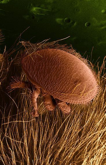
| 22:04, 19 May 2010 | Varroa logo.jpg (file) | 71 KB | Image sourced from Wikimedia Commons. This image is in the public domain because it contains materials that originally came from the Agricultural Research Service, the research agency of the United States Department of Agriculture. | 1 | |
| 21:01, 18 May 2015 | Vapenec transparent.png (file) | 156 KB | 1 | ||
| 12:44, 27 April 2012 | VPH.png (file) |  |
21 KB | update | 2 |
| 22:55, 12 December 2009 | Uterine tumour.jpg (file) |  |
6 KB | Courtesy of The Donkey Sanctuary | 1 |
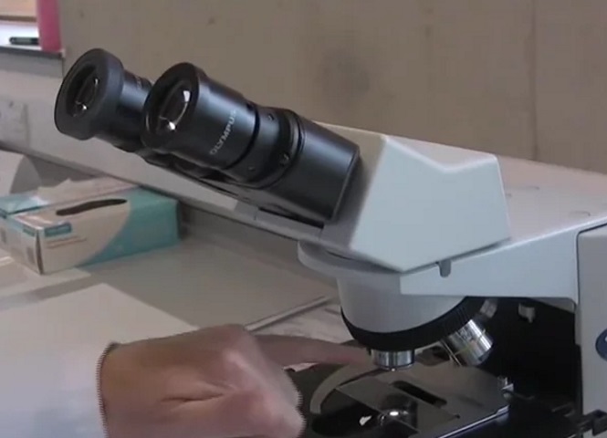
| 15:16, 27 September 2016 | Using a microscope.jpg (file) |  |
51 KB | video screenshot | 1 |
| 15:39, 28 September 2011 | Urinary.png (file) |  |
20 KB | {{Information |Description=Button for Urinary System |Source=Own work |Date=28 September 2011 |Author=User:Bara |Permission=See below |other_versions= }} | 1 |
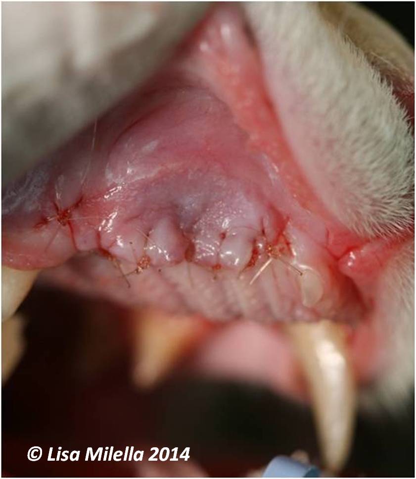
| 14:25, 1 May 2014 | Upper Canine Extraction 5.jpg (file) |  |
55 KB | {{Information |Description ={{en|1=The flap is replaced and sutured in position using a monofilament absorbable suture material, using a simple interrupted suture pattern. }} |Source =Lisa Milella |Author =Lisa Milella |Date = | 1 |
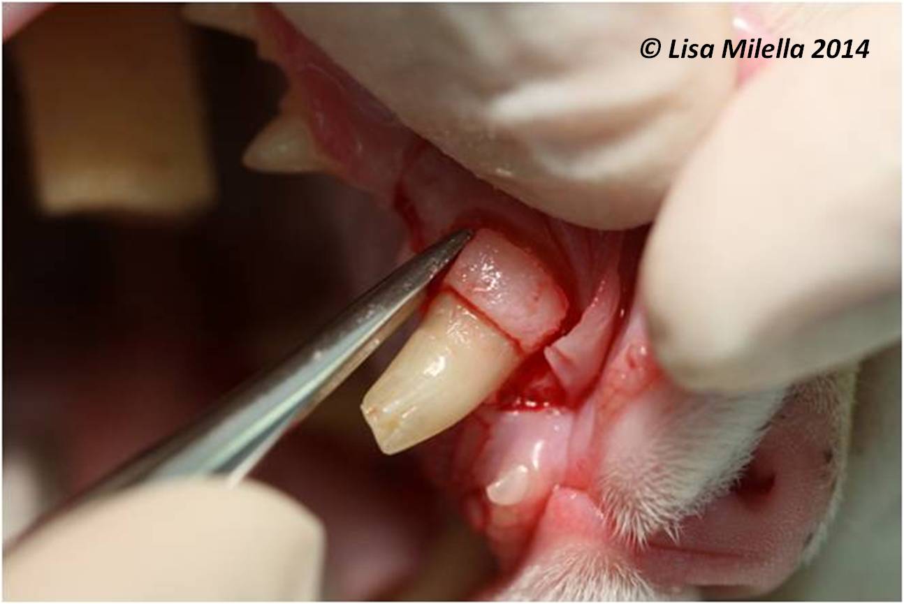
| 14:22, 1 May 2014 | Upper Canine Extraction 4.jpg (file) |  |
70 KB | {{Information |Description ={{en|1=When the tooth is loose, position the extraction forceps as far apically as possible and rotate the tooth along its long axis, pulling gently at the same time.}} |Source =Lisa Milella |Author =Lisa Mil | 1 |
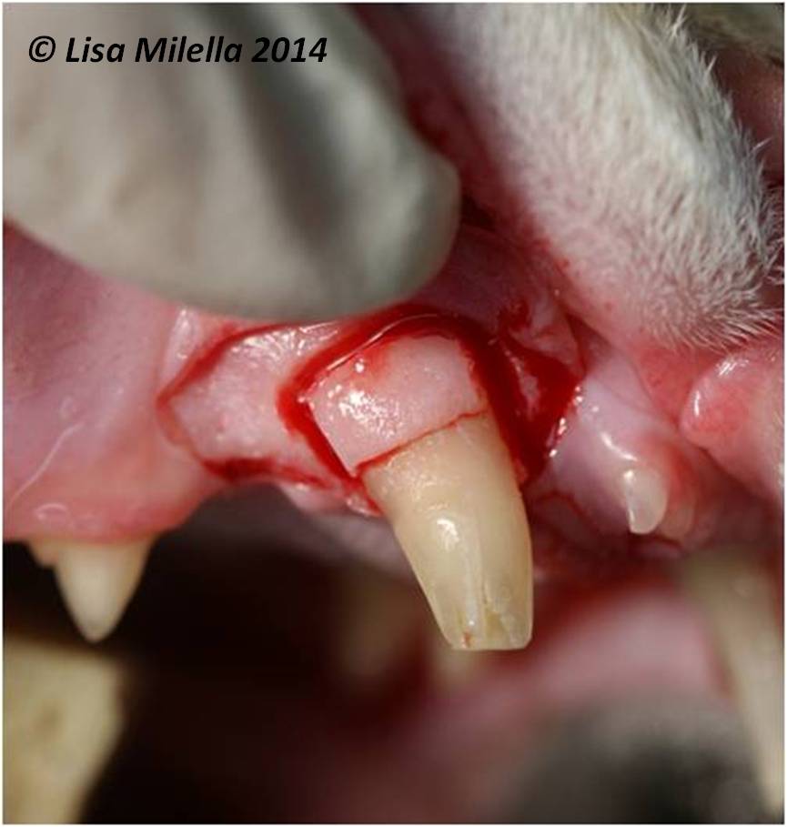
| 14:17, 1 May 2014 | Upper Canine Extraction 3.jpg (file) |  |
64 KB | {{Information |Description ={{en|1=A dental elevator is positioned in the groove created on either the mesial or distal aspect of the tooth. }} |Source =Lisa Milella |Author =Lisa Milella |Date =2014 |Permission = |other_v | 1 |
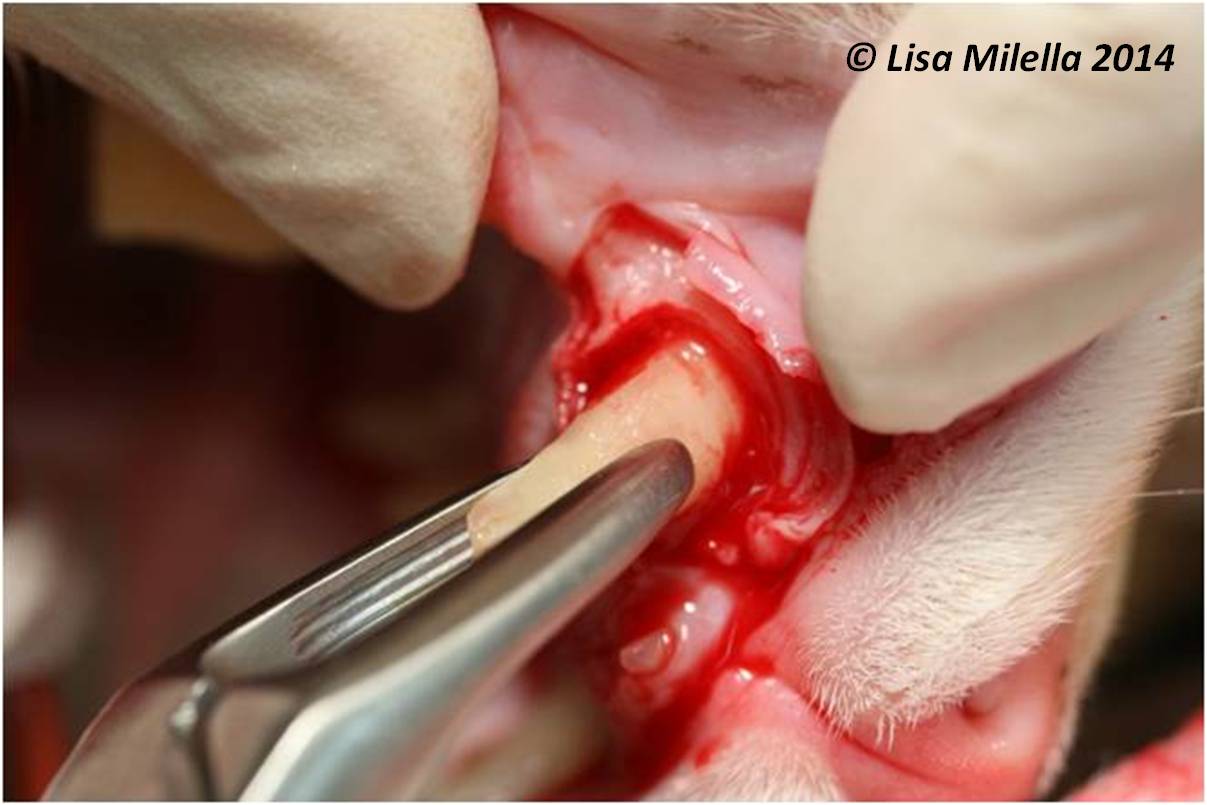
| 14:12, 1 May 2014 | Upper Canine Extraction 2.jpg (file) |  |
53 KB | {{Information |Description ={{en|1=Some overlying buccal bone can be removed to adequately see the mesial and distal edge of the tooth root. The gutters should be half the width of the tooth root and extend up to 2/3 of the length of the root. The gutt | 1 |