Uploads by Ctrace
This special page shows all uploaded files.
| Date | Name | Thumbnail | Size | Description | Versions |
|---|---|---|---|---|---|
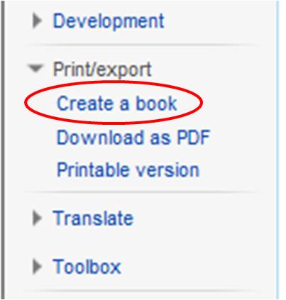
| 14:59, 19 December 2011 | Createabook2.jpg (file) |  |
16 KB | Updated image showing user's skin | 2 |
| 17:21, 12 December 2011 | PinR.png (file) |  |
844 bytes | Made smaller, to 15x27 pixels | 2 |
| 17:18, 12 December 2011 | PinB.png (file) |  |
927 bytes | Made 16x28 pixels | 3 |
| 17:06, 12 December 2011 | PinBD.png (file) |  |
1 KB | Changed pin so has no dot in the middle | 2 |
| 18:03, 24 November 2011 | StarPin.png (file) |  |
862 bytes | {{Information |Description ={{en|1=Star that goes in pin on Vet schools worldwide map, used to denote a school that has a student ambassador}} |Source =Own work |Author =Ctrace |Date =24/11/2011 |Permission | 1 |
| 17:59, 24 November 2011 | YellowPin.png (file) |  |
1 KB | {{Information |Description ={{en|1=Yellow pin to denote a school that has a school page}} |Source =Own work |Author =Ctrace |Date = |Permission = |other_versions = }} | 1 |
| 17:58, 24 November 2011 | RedPin.png (file) |  |
1 KB | Pin used for Vetschools world map to denote a school without a page in WikiVet | 1 |
| 15:36, 21 November 2011 | WikiVet Newsletter Issue 8.pdf (file) | 275 KB | 8th Issue of the WikiVet Newsletter, PDF version | 1 | |
| 11:08, 18 November 2011 | WikiVet Newsletter Issue 9.pdf (file) | 282 KB | {{Information |Description ={{en|1=WikiVet Newsletter Issue 9, November 2011}} |Source =Own work |Author =Ctrace |Date =16/11/2011 |Permission = |other_versions = }} | 1 | |
| 12:42, 15 November 2011 | Aspark.png (file) |  |
115 KB | {{Information |Description ={{en|1=cropped version of original image}} |Source =File:155003 1630342032382 1051549747 2555529 2021648 n (1).jpg |Author =Alistair Spark |Date =15/11/2011 |Permission = |other_versions = | 1 |
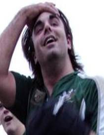
| 12:36, 15 November 2011 | Aspark.jpg (file) |  |
7 KB | {{Information |Description ={{en|1=Cropped version of original image}} |Source =File:155003 1630342032382 1051549747 2555529 2021648 n (1).jpg |Author =Alistair Spark |Date =15/11/2011 |Permission = |other_versions = | 1 |
| 11:27, 15 November 2011 | OER.jpg (file) |  |
5 KB | {{Information |Description ={{en|1=OER (Open Educational Resources) logo}} |Source =OER |Author =OER |Date = |Permission = |other_versions = }} | 1 |
| 11:41, 14 November 2011 | WikiCoursessmall.png (file) |  |
10 KB | {{Information |Description ={{en|1=Smaller version of the WikiVet courses logo, 100px wide}} |Source =Own work |Author =Ctrace |Date =14/11/2011 |Permission = |other_versions = }} | 1 |
| 11:36, 14 November 2011 | WikiCourses.png (file) |  |
84 KB | {{Information |Description ={{en|1=Logo for WikiVet courses}} |Source =Own work |Author =Ctrace |Date =14/11/2011 |Permission = |other_versions = }} | 1 |
| 11:24, 14 November 2011 | PowerPoint.png (file) |  |
108 KB | WV logo's aligned a bit better | 2 |
| 10:10, 14 November 2011 | WVpodcastssmall.png (file) |  |
12 KB | {{Information |Description ={{en|1=Small version of the WikiVet Podcast logo}} |Source =Own work |Author =Ctrace |Date =10/11/2011 |Permission = |other_versions = }} | 1 |
| 10:07, 14 November 2011 | WVpodcasts.png (file) |  |
123 KB | Logo image for WikiVet podcasts | 1 |
| 09:24, 19 September 2011 | Vetimpulse.jpg (file) |  |
4 KB | {{Information |Description ={{en|1=Screenshot of Vet impulse logo taken from their website with permission}} |Source =http://www.vet-impulse.com/ |Author =http://www.vet-impulse.com/ |Date =19/09/2011 |Permission = |other_ | 1 |
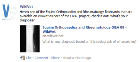
| 09:01, 19 September 2011 | Facebooksharelink.png (file) |  |
51 KB | {{Information |Description ={{en|1=Screenshot of WikiVet's facebook page, cropped to show posted link with the 'share' link.}} |Source =http://www.facebook.com/pages/WikiVet/188576847835536 |Author =Ctrace |Date | 1 |
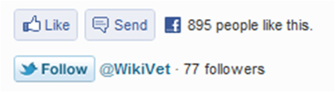
| 08:40, 19 September 2011 | Facebooksendlink.png (file) |  |
38 KB | {{Information |Description ={{en|1=Screenshot of the WikiVet homepage, cropped to show the Facebook like and send buttons and the Twitter follow button}} |Source =WikiVet home page |Author =Ctrace |Date =19/09/ | 1 |
| 15:16, 14 September 2011 | WikiVetneedsyou.png (file) |  |
2.38 MB | {{Information |Description ={{en|1=WikiVet logo with 'your school needs YOU!' text}} |Source =Own work |Author =Ctrace and Brian Cox |Date =13th September 2011 |Permission = |other_versions = }} | 1 |
| 10:47, 7 September 2011 | WikiVet Newsletter issue 7.pdf (file) | 246 KB | {{Information |Description =7th Issue of the WikiVet newsletter |Source =WikiVet |Author =Chris Trace |Date =Sent 21st July 2011 |Permission = |other_versions = }} | 1 | |
| 17:12, 15 July 2011 | VPU.jpg (file) |  |
13 KB | {{Information |Description ={{en|1=Logo for the Veterinary Pathology Unit, Royal (Dick) School of Veterinary Studies, University of Edinburgh}} |Source =Veterinary Pathology Unit, Royal (Dick) School of Veterinary Studies, University of Edinbur | 1 |
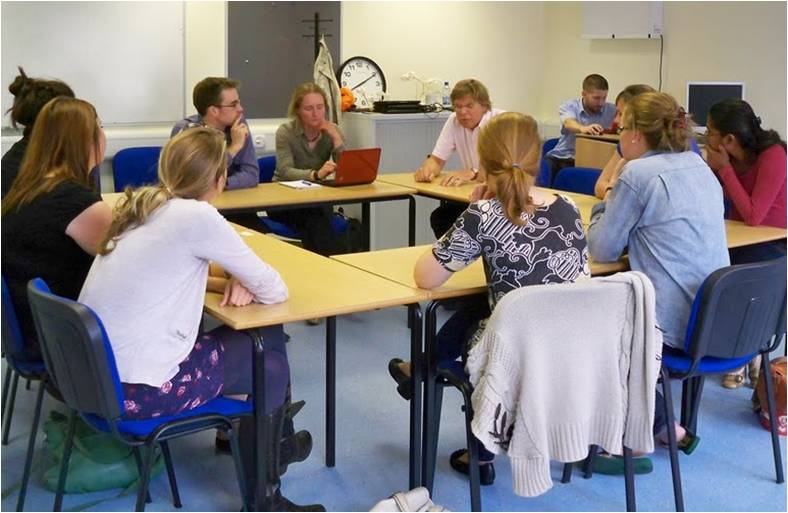
| 16:29, 15 July 2011 | Students.JPG (file) |  |
57 KB | {{Information |Description ={{en|1=Picture of Student focus group at the 2nd Annual Veterinary Educational Symposium in Nottingham}} |Source =Own work |Author =Ctrace |Date =12th July 2011 |Permission = |ot | 1 |
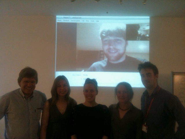
| 16:06, 15 July 2011 | GlasgowTELT.jpg (file) |  |
82 KB | {{Information |Description ={{en|1=WikiVet group at TELT day in Glasgow Vet school. From left to right: Nick Short, Ute Barrett, Glasgow Student, Barbora Stanikova, Chris Trace, Background shows another Glasgow student in Canada via Skype}} |Source | 1 |
| 14:54, 11 July 2011 | Right atrium.jpg (file) |  |
24 KB | {{Information |Description ={{en|1=Image of right atrium of heart - see [http://en.wikivet.net/R(D)SVS_VPU_01 case in WikiVet]}} |Source =Veterinary Pathology Unit, Royal (Dick) School of Veterinary Studies, University of Edinburgh |Author | 1 |
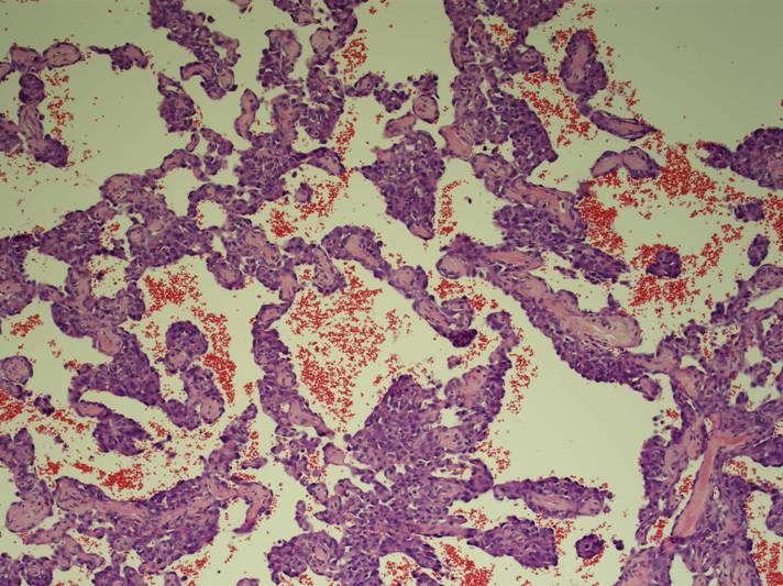
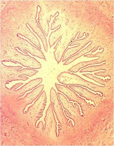
| 14:52, 11 July 2011 | Low power.jpg (file) |  |
92 KB | {{Information |Description ={{en|1=Low-power micrograph image of a Haemangiosarcoma}} |Source =Veterinary Pathology Unit, Royal (Dick) School of Veterinary Studies, University of Edinburgh |Author =Chris Palgrave |Date =2010 | | 1 |
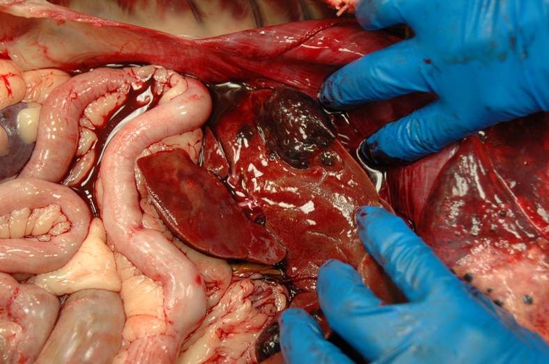
| 14:50, 11 July 2011 | Liver pathology.jpg (file) |  |
64 KB | {{Information |Description ={{en|1=Masses in liver of a dog}} |Source =Veterinary Pathology Unit, Royal (Dick) School of Veterinary Studies, University of Edinburgh |Author =Chris Palgrave |Date =2010 |Permission = |other_ | 1 |
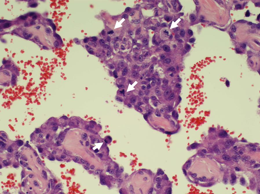
| 14:47, 11 July 2011 | High power.jpg (file) |  |
89 KB | {{Information |Description ={{en|1=Micrograph of a haemangiosarcoma, with mitotic figures labelled}} |Source =Veterinary Pathology Unit, Royal (Dick) School of Veterinary Studies, University of Edinburgh |Author =Chris Palgrave |Date | 1 |
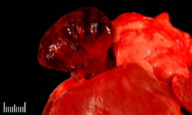
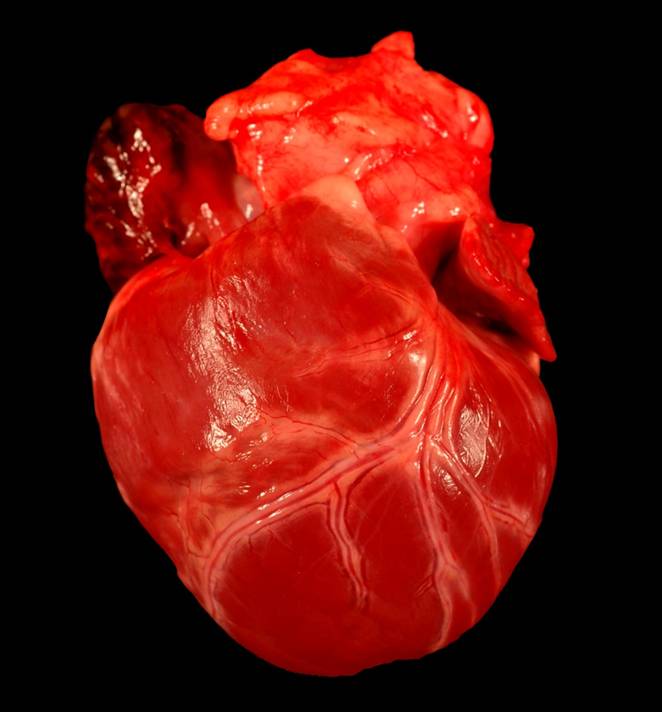
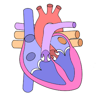
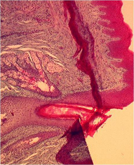
| 14:42, 11 July 2011 | Gross Heart.jpg (file) |  |
37 KB | {{Information |Description ={{en|1=Image of a heart of a dog - see [http://en.wikivet.net/R(D)SVS_VPU_01 case in WikiVet]}} |Source =Veterinary Pathology Unit, Royal (Dick) School of Veterinary Studies, University of Edinburgh |Author = | 1 |
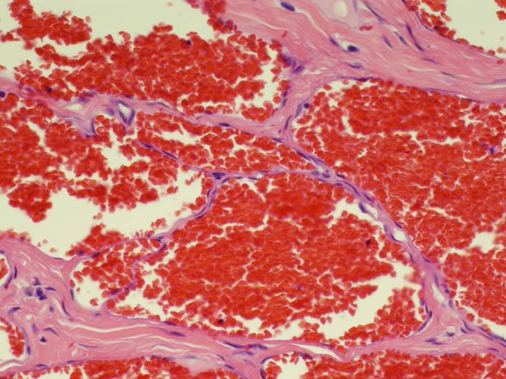
| 14:33, 11 July 2011 | Haemangioma.jpg (file) |  |
74 KB | {{Information |Description ={{en|1=A micrograph of a haemangioma}} |Source =Veterinary Pathology Unit, Royal (Dick) School of Veterinary Studies, University of Edinburgh |Author =Chris Palgrave |Date =2010 |Permission = |o | 1 |
| 11:08, 1 July 2011 | Dragster logo.png (file) | 26 KB | {{Information |Description=Blue jigsaw piece, dragster logo with transparent background |Source=RVC |Date=File Originally created 19/05/2011, re-uploaded in png format with transparent background 1/0 | 1 | |
| 12:38, 26 June 2011 | Cardiovascular logo.png (file) | 42 KB | Cropped version for use in frontpage templates | 2 | |
| 12:30, 20 June 2011 | VetEdLogo.jpg (file) |  |
54 KB | {{Information |Description=Logo for Nottingham 2011 Veterinary Educational Symposium |Source=Royal Veterinary College |Date=15th June 2011 |Author=Tierney Kinnison |Permission= |other_versions= }} | 1 |
| 10:01, 16 June 2011 | WikiVet Newsletter Issue 6.pdf (file) | 246 KB | {{Information |Description =6th Issue of the WikiVet newsletter |Source =WikiVet |Author =Chris Trace |Date =Sent 10th June 2011 |Permission = |other_versions = }} | 1 | |
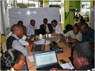
| 15:59, 9 June 2011 | WikiVet Africa.jpg (file) |  |
19 KB | {{Information |Description =Image of staff at a Kenyan vet school exploring WikiVet |Source =Nick Short |Author =Nick Short |Date =May 2011 |Permission = |other_versions = }} | 1 |
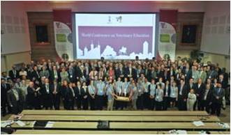
| 11:40, 9 June 2011 | Vet2011 group.jpg (file) |  |
15 KB | {{Information |Description =Group photo of delegates at the Vet2011 World conference on Veterinary education |Source =Julien Durand |Author =Julien Durand |Date =14th May 2011 - uploaded to WikiVet 9th June 2011 |Permission | 1 |
| 14:55, 6 June 2011 | Pituitary.jpg (file) |  |
39 KB | {{Information |Description=Screenshot from Powerpoint, courtesy of the Royal Veterinary College. Shows histological section of the pituitary gland. |Source=The Royal Veterinary College |Date=06/06/2011 |Au | 1 |
| 14:47, 6 June 2011 | Endocrine.swf (file) | 1.98 MB | {{Information |Description=PowerPoint tutorial on Endocrine histology, converted to swf format. Resource page can be found here |Source=The Royal Veterinary College |Date=2011 |Author=[[RVC|The Royal Veterinary Co | 1 | |
| 13:27, 6 June 2011 | Spermatazoa.jpg (file) |  |
28 KB | {{Information |Description=Screenshot from Powerpoint, courtesy of the Royal Veterinary College. Shows scanning electron micrograph of a spermatazoan. |Source=The Royal Veterinary College |Da | 1 |
| 12:24, 6 June 2011 | Male Reproductive Tract.swf (file) | 1.87 MB | {{Information |Description=PowerPoint tutorial on Male reproductive tract histology, converted to swf format. Resource page can be found here |Source=The Royal Veterinary College |Date=2011 |Author=[ | 1 | |
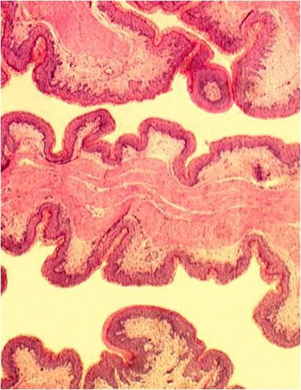
| 11:18, 6 June 2011 | Cervix.jpg (file) |  |
43 KB | {{Information |Description=Screenshot from Powerpoint, courtesy of the Royal Veterinary College. Shows transverse histological section through cervix. |Source=The Royal Veterinary College | | 1 |
| 11:12, 6 June 2011 | Female Reproductive Tract.swf (file) | 1.98 MB | {{Information |Description=PowerPoint tutorial on Female (non-pregnant) reproductive tract histology, converted to swf format. Resource page can be found here |Source=The Royal Veterinary College | | 1 | |
| 15:54, 3 June 2011 | Mammary.jpg (file) |  |
55 KB | {{Information |Description=Screenshot from Powerpoint, courtesy of the Royal Veterinary College. Shows teat canal. |Source=The Royal Veterinary College |Date=03/06/2011 |Author=Chris Trace |Permission= | 1 |
| 15:49, 3 June 2011 | Mammary Gland.swf (file) | 1.93 MB | {{Information |Description=PowerPoint tutorial on Mammary Gland histology, converted to swf format. Resource page can be found here |Source=The Royal Veterinary College |Date=2011 |Author=[[RVC|The Royal Veter | 1 | |
| 14:57, 3 June 2011 | GIT-3.jpg (file) |  |
47 KB | {{Information |Description=Screenshot from Powerpoint, courtesy of the Royal Veterinary College. Shows folds of laminae in the abomasum. |Source=The Royal Veterinary College |Date=03/ | 1 |
| 14:52, 3 June 2011 | GIT-3.swf (file) | 1.98 MB | {{Information |Description=PowerPoint tutorial on Ruminant Gastrointestinal tract histology, converted to swf format. Resource page can be found here |Source=The Royal Veterinary College |Dat | 1 | |
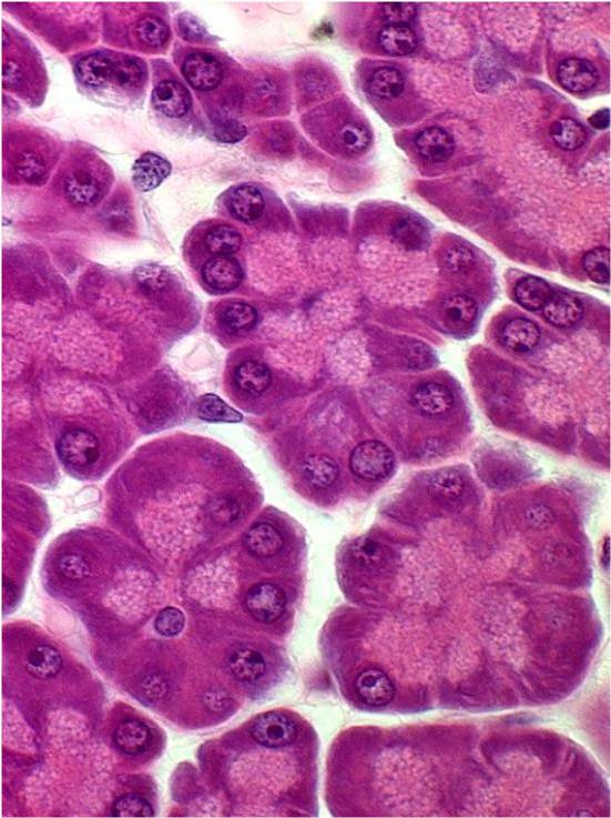
| 12:49, 3 June 2011 | GIT-2.jpg (file) |  |
72 KB | {{Information |Description=Screenshot from GIT part 2 Powerpoint, courtesy of the Royal Veterinary College. Shows exocrine acini of pancreas. |Source=The Royal Veterinary College |Date=03/06/2 | 1 |
| 12:43, 3 June 2011 | GIT-2.swf (file) | 2 MB | {{Information |Description=PowerPoint tutorial on Gastrointestinal tract histology, converted to swf format. Resource page can be found here |Source=The Royal Veterinary College |Date=2011 |Author=[[R | 1 | |
| 11:32, 3 June 2011 | GIT-1.jpg (file) |  |
41 KB | {{Information |Description=Screenshot from GIT part 1 Powerpoint, courtesy of the Royal Veterinary College. Shows transverse section through colon. |Source=The Royal Veterinary College |Date=0 | 1 |
.jpg)