Uploads by Lizzies
This special page shows all uploaded files.
| Date | Name | Thumbnail | Size | Description | Versions |
|---|---|---|---|---|---|
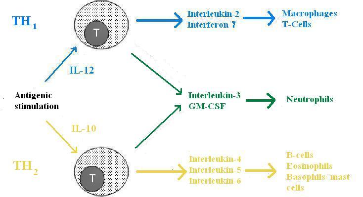
| 16:44, 5 September 2007 | TH function.jpg (file) |  |
33 KB | Functions of TH cells. Pending permission from John Hopkins. | 1 |
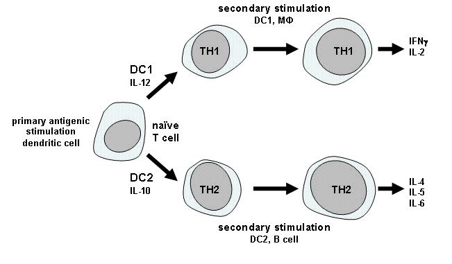
| 16:24, 5 September 2007 | TH development.jpg (file) |  |
32 KB | Development of the TH subpopulations. Pending permission from John Hopkins. | 1 |
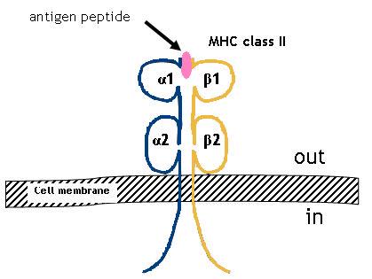
| 09:57, 5 September 2007 | MHC II.jpg (file) |  |
19 KB | Structure of MHC II. Pending permission from John Hopkins. | 1 |
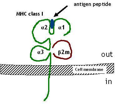
| 09:29, 5 September 2007 | MHC I.jpg (file) |  |
19 KB | Structure of MHC class I. Pending permission from John Hopkins. | 1 |
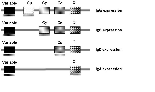
| 16:53, 4 September 2007 | Immunoglobulin heavy chain genes.jpg (file) |  |
29 KB | Immunoglobulin heavy chain genes. Pending permission from John Hopkins. | 1 |
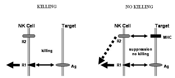
| 16:27, 4 September 2007 | NK cell killing.jpg (file) |  |
16 KB | Diagrammatic representation of the receptors involved in NK cell killing. Pending permission from John Hopkins. | 1 |
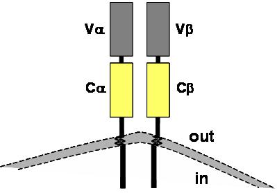
| 16:10, 4 September 2007 | TcR structure.jpg (file) |  |
11 KB | Diagram showing the basic structure of a TcR. Pending permission from John Hopkins. | 1 |
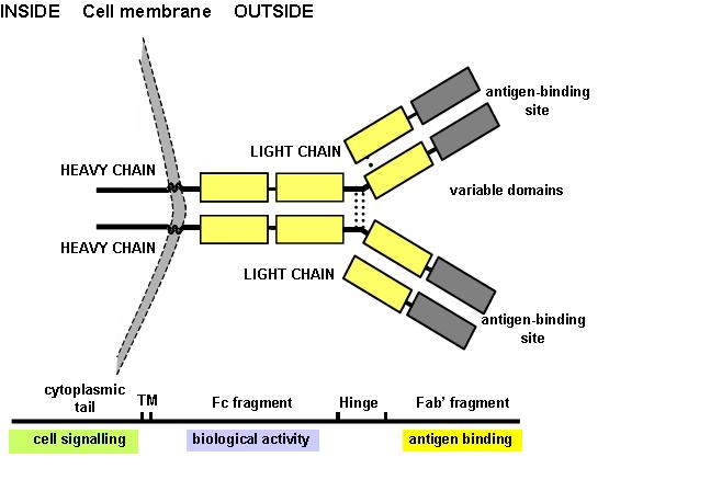
| 16:04, 4 September 2007 | Antibody structure.jpg (file) |  |
34 KB | Diagram showing the basic structure of antibody. Pending permission from John Hopkins. | 1 |
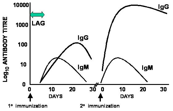
| 15:46, 4 September 2007 | Primary and secondary response.jpg (file) |  |
25 KB | Time course of the primary and secondary responses. Pending permission from John Hopkins. | 1 |
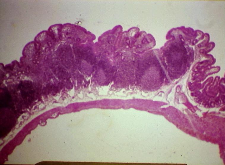
| 08:33, 4 September 2007 | Peyers patches.jpg (file) |  |
57 KB | Peyer's patches. Courtesy of BioMed Archive. | 1 |
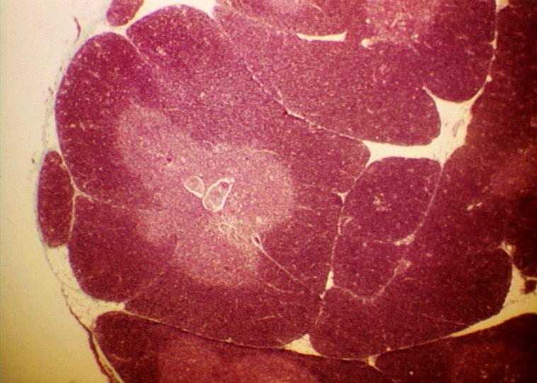
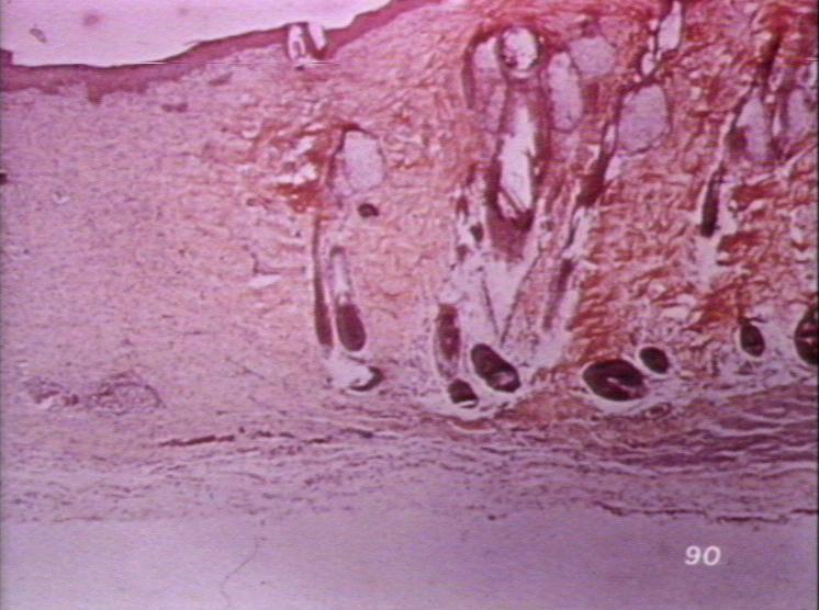
| 16:59, 3 September 2007 | Thymus histo.jpg (file) |  |
68 KB | Histological appearance of the thymus. Courtesy of BioMed Archive. | 1 |
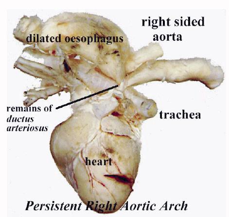
| 15:29, 3 September 2007 | Persistent right aortic arch.jpg (file) |  |
32 KB | Persistent right aortic arch. Pending permission from Brian Smyth. | 1 |
| 15:28, 3 September 2007 | Hydrocephalus.jpg (file) |  |
67 KB | Hydrocephalus in the puppy. Pending permission from Brian Smyth. | 1 |
| 15:27, 3 September 2007 | Cleft palate.jpg (file) |  |
21 KB | Cleft palate in the cat. Pending permission from Brian Smyth. | 1 |
| 14:21, 3 September 2007 | Cyclops.jpg (file) |  |
35 KB | Cyclops. Pending permission from Brian Smyth. | 1 |
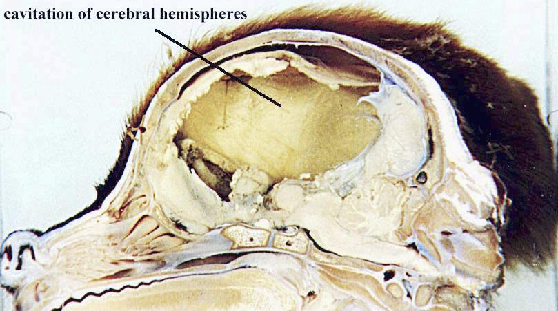
| 13:59, 3 September 2007 | Swayback sections.jpg (file) |  |
40 KB | Sectioned brain showing "swayback". There is extensive lysis of subcortical white matter lateral to the lateral ventricle of the cerebrum. Pending permission from Brian Smyth. | 1 |
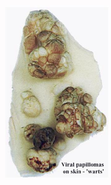
| 13:19, 3 September 2007 | Viral papilloma.jpg (file) |  |
27 KB | Viral papillomas on the skin. Pending permission from Brian Smyth. | 1 |
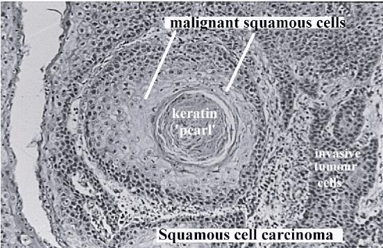
| 13:17, 3 September 2007 | Squamous cell carcinoma histo.jpg (file) |  |
61 KB | Histological appearance of squamous cell carcinoma. Pending permissions from Brian Smyth. | 1 |
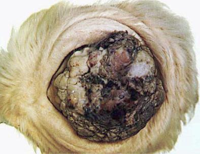
| 13:17, 3 September 2007 | Squamous cell carcinoma eye.jpg (file) |  |
23 KB | Squamous cell carcinoma of the eye of a hereford cow. UV light contributes to causing the tumour. Pending permission from Brian Smyth. | 1 |
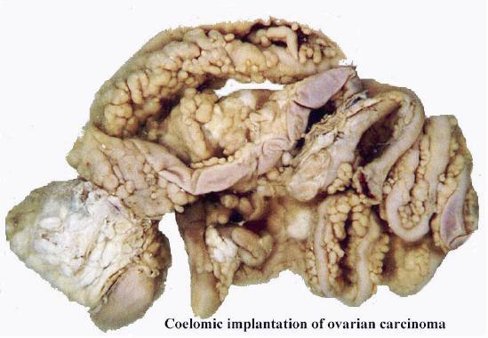
| 11:56, 3 September 2007 | Coelomic implantation ovarian carcinoma.jpg (file) |  |
36 KB | Coelomic implantation of ovarian carcinoma. Pending permission from Brian Smyth. | 1 |
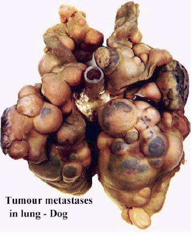
| 11:55, 3 September 2007 | Tumour metastases lung dog.jpg (file) |  |
30 KB | Tumour metastases in the lung of the dog. Pending permission from Brian Smyth. | 1 |
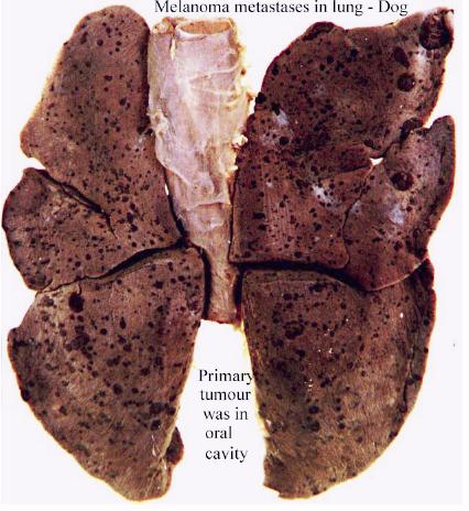
| 11:54, 3 September 2007 | Melanoma metastases dog lung.jpg (file) |  |
44 KB | Lung metastases of melanoma in the dog. Pending permission from Brian Smyth. | 1 |
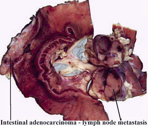
| 11:54, 3 September 2007 | Intestinal adenocarcinoma lymphatic spread.jpg (file) |  |
40 KB | Gross appearance of lymphatic spread of intestinal adenocarcinoma. Pending permission from Brian Smyth. | 1 |
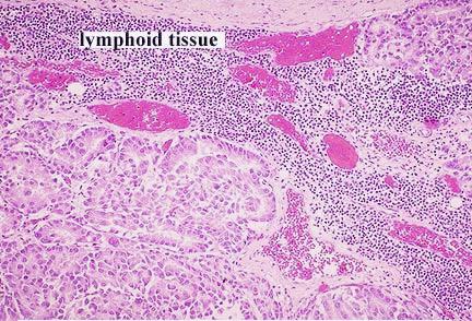
| 11:53, 3 September 2007 | Adenocarcinoma metastasis to lymph node.jpg (file) |  |
52 KB | Histological appearance of lymphatic spread of adenocarcinoma to the lymph nodes. Pending permission from Brian Smyth. | 1 |
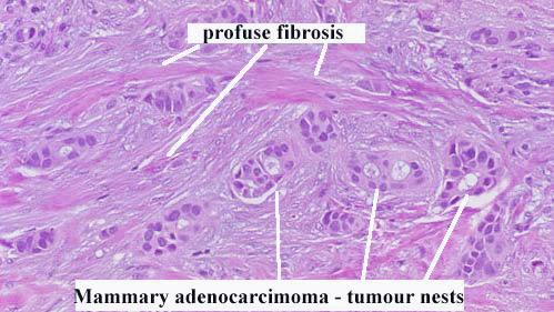
| 11:44, 3 September 2007 | Profuse fibrosis.jpg (file) |  |
37 KB | Profuse fibrosis in mammary adenocarcinoma. Pending permission from Brian Smyth. | 1 |
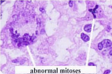
| 11:42, 3 September 2007 | Abnormal mitoses.jpg (file) |  |
17 KB | Abnormal mitoses. Pending permission from Brian Smyth. | 1 |
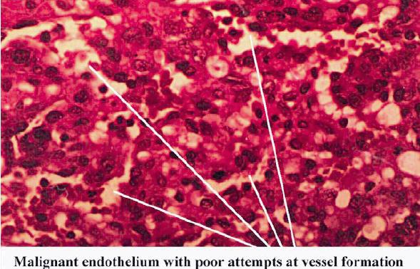
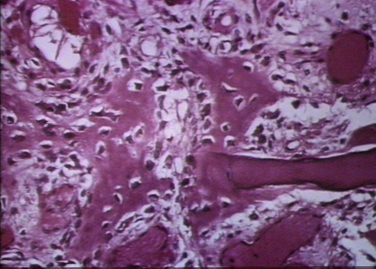
| 11:36, 3 September 2007 | Haemangioscarcoma histo.jpg (file) |  |
58 KB | Histological appearance of malignant endothelium. Malignant tumours tend to appear more anaplastic and basophilic, and have a higher nucleus:cytoplasm ratio. Pending permission from Brian Smyth. | 1 |
| 11:32, 3 September 2007 | Haemangioscarcoma lung metastases.jpg (file) |  |
38 KB | Metastases in the lung of the dog, from haemangiosarcoma of the flank. Malignant tumours can, and some readily do, spread to local lymph nodes and the lungs. Pending permssion from Brian Smyth. | 1 |
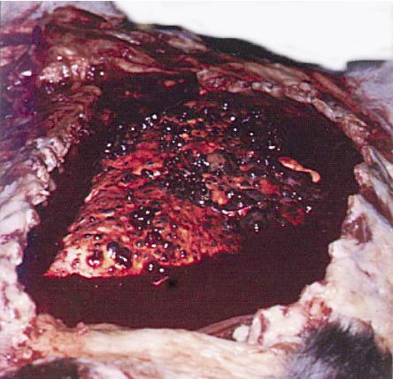
| 11:31, 3 September 2007 | Haemangioscarcoma flank.jpg (file) |  |
57 KB | Haemangiosarcoma (malignant) in the flank of a dog. Pending permission from Brian Smyth. | 1 |
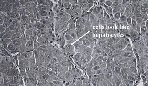
| 11:17, 3 September 2007 | Perianal gland hepatoid adenoma dog.jpg (file) |  |
37 KB | Perianal gland "hepatoid" adenoma in the dog (benign). Note the well differentiated cells, that resemble hepatocytes. The tissue is well organised, there are few mitoses and there is no haemoorhage or necrosis. Pending permission from Brian Smyth. | 1 |
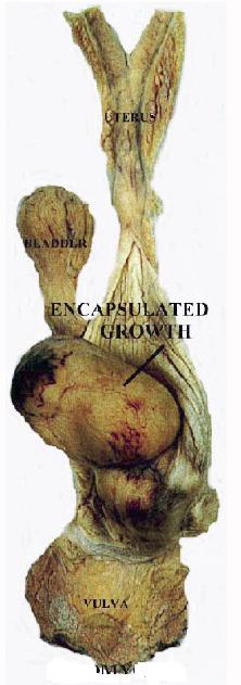
| 11:14, 3 September 2007 | Benign leiomyoma.jpg (file) |  |
25 KB | Benign neoplasm (leiomyoma). Note that the growth is encapsulated. Pending permission from Brian Smyth. | 1 |
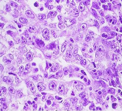
| 12:21, 31 August 2007 | Anaplatic carcinoma.jpg (file) |  |
41 KB | Anaplastic carcinoma. Pending permission from Brian Smyth. | 1 |
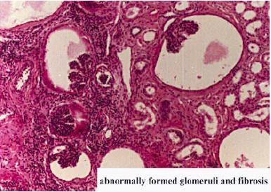
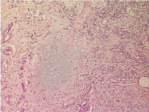
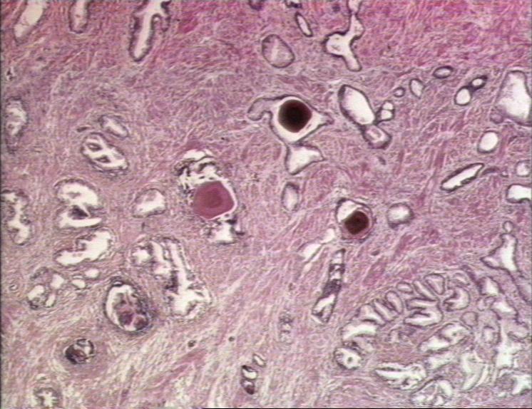
| 12:18, 31 August 2007 | Renal dysplasia dog histological.jpg (file) |  |
31 KB | Histological appearance of renal dysplasia in the dog. Note the abnormally formed glomeruli and fibrosis. Pending permission from Brian Smyth. | 1 |
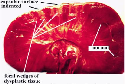
| 12:18, 31 August 2007 | Renal dysplasia dog gross.jpg (file) |  |
24 KB | Gross apperarance of renal dysplasia in the dog. Pending permission from Brian Smyth. | 1 |
| 12:13, 31 August 2007 | Mammary tumour ossification.jpg (file) |  |
59 KB | Bone formation in a mixed mammary tumour. Pending permission from Brian Smyth. | 1 |
| 12:04, 31 August 2007 | Benign prostatic hyperplasia.jpg (file) |  |
89 KB | Benign prostatic hyperplasia (man). Courtesy of BioMed Archive. | 1 |
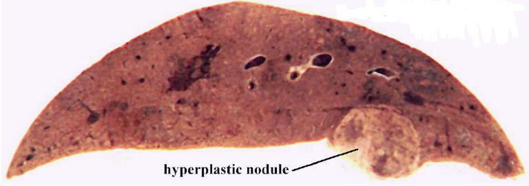
| 12:00, 31 August 2007 | Hyperplastic nodule liver.jpg (file) |  |
26 KB | Hyperplastic nodule in the liver of an elderly dog. Pending permission from Brian Smyth. | 1 |
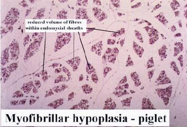
| 11:50, 31 August 2007 | Myofibrillar hypoplasia.jpg (file) |  |
25 KB | Myofibrillar hypoplasia in the piglet. Pending permission from Brian Smyth. | 1 |
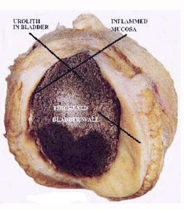
| 11:45, 31 August 2007 | Obstructional hypertrophy bladder.jpg (file) |  |
27 KB | Hypertrophy of the bladder wall in response to a urolith (urinary calculus; bladder stone). Pending permission from Brian Smyth. | 1 |
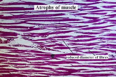
| 11:35, 31 August 2007 | Muscle atrophy.jpg (file) |  |
31 KB | Histological appearance of muscle atrophy. Pending permission from Brian Smyth. | 1 |
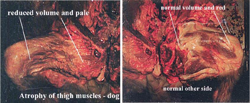
| 11:32, 31 August 2007 | Thigh muscles atrophy.jpg (file) |  |
71 KB | Atrophy of the thigh muscles of the dog. The left hand side shows the atrophied muscles, whereas the right hand side shows the normal limb on the other side. Pending permission from Brian Smyth. | 1 |
| 10:51, 31 August 2007 | Scar tissue.jpg (file) |  |
64 KB | Histological appearance of scar tissue. Courtesy of BioMed Archive. | 1 |
| 10:48, 31 August 2007 | Fracture repair.jpg (file) |  |
70 KB | Histological appearance of a repairing fracture. Courtesy of BioMed Archive. | 1 |
| 10:46, 31 August 2007 | Fracture callus.jpg (file) |  |
72 KB | Fracture callus. Courtesy of BioMed Archive. | 1 |
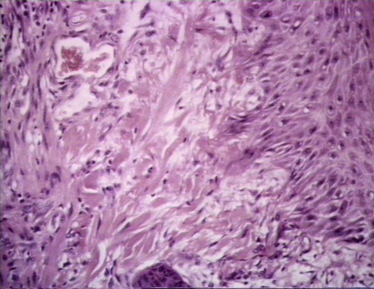
| 10:44, 31 August 2007 | Granultion tissue histology.jpg (file) |  |
74 KB | Histological appearance of maturing granulation tissue. Courtesty of BioMed Archive. | 1 |
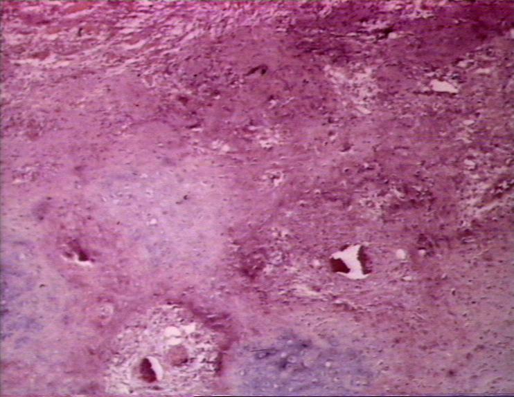
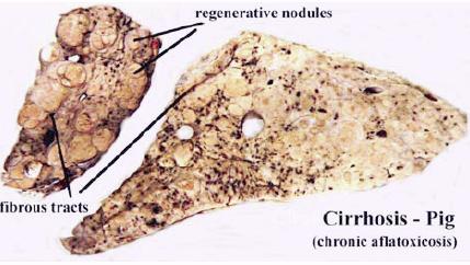
| 10:31, 31 August 2007 | Cirrhosis pig.jpg (file) |  |
25 KB | Cirrhosis, due to chronic liver damage in the pig. Pending permission fro Brian Smyth. | 1 |
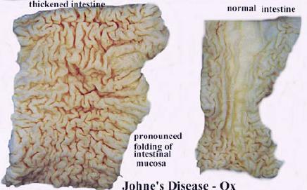
| 10:25, 31 August 2007 | Johnes disease comparative.jpg (file) |  |
23 KB | Comparative gross appearance of Johne's Disease in the ox. The intestine on the right is diseased, whereas that on the left is normal. Pending permission from Brian Smyth. | 1 |
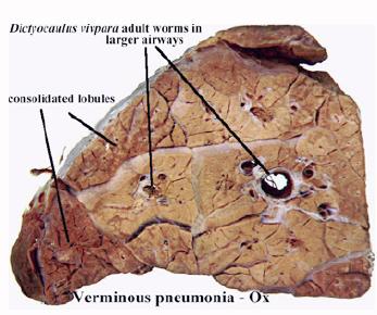
| 10:20, 31 August 2007 | Lungworm.jpg (file) |  |
24 KB | ''Dictyocaulus vivipara'' in the lung of the ox. Causes verminous pneumonia. Pending permission from Brian Smyth. | 1 |
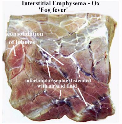
| 10:14, 31 August 2007 | Fog fever.jpg (file) |  |
33 KB | Gross appearance of "Fog Fever" in the ox. Pending permission from Brian Smyth. | 1 |
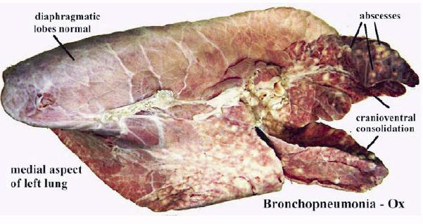
| 10:11, 31 August 2007 | Bronchopneumonia ox.jpg (file) |  |
40 KB | Bronchonpneumonia in the ox (gross). Pending permission from Brian Smyth. | 1 |