File list
This special page shows all uploaded files.
| Date | Name | Thumbnail | Size | User | Description | Versions |
|---|---|---|---|---|---|---|
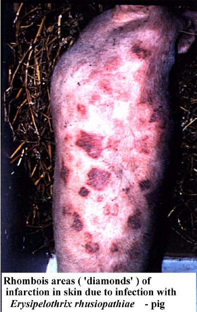
| 12:20, 29 August 2007 | Skin infarction pig.jpg (file) |  |
51 KB | Lizzies | Infarction in the skin of the pig caused by ''Erysipelothrix rhusiopathiae''. Pending permission from Brian Smyth. | 1 |
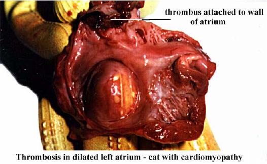
| 12:09, 29 August 2007 | Thrombus cat left atrium.jpg (file) |  |
31 KB | Lizzies | Gross appearance of a thrombus attached to the wall of the left atrium in a cat suffering from cardiomyopathy. Pending permission from Brian Smyth. | 1 |
| 15:32, 28 August 2007 | VSD1.jpg (file) |  |
90 KB | Kjr35 | 1 | |
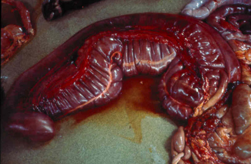
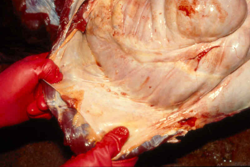
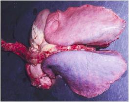
| 09:31, 28 August 2007 | Large colon torsion horse.jpg (file) |  |
110 KB | Mayazoey | Large colon torsion in the horse. Courtesy of Elspeth Milne, University of Edinburgh. | 1 |
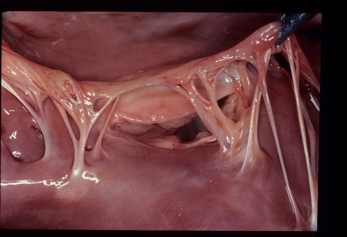
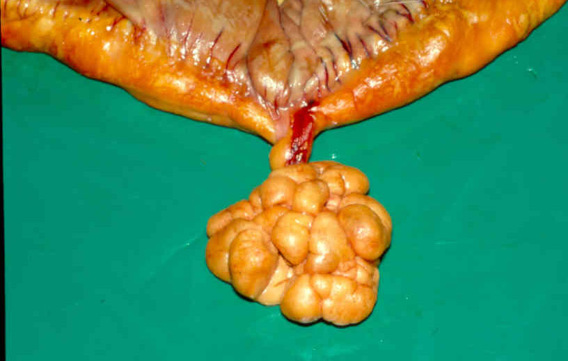
| 09:29, 28 August 2007 | Pedunc lipoma closeup.jpg (file) |  |
71 KB | Mayazoey | Pedunculated lipoma in the horse. Courtesy of Elspeth Milne, University of Edinburgh. | 1 |
| 09:23, 28 August 2007 | Colon adhesions horse.jpg (file) |  |
88 KB | Mayazoey | Colonic adhesions in the horse. Courtesy of Elspeth Milne, University of Edinburgh. | 1 |
| 09:23, 22 August 2007 | Cocktails.jpg (file) |  |
30 KB | Lizzies | 1 | |
| 08:50, 22 August 2007 | My dog and I.jpeg (file) |  |
73 KB | Bara | 1 | |
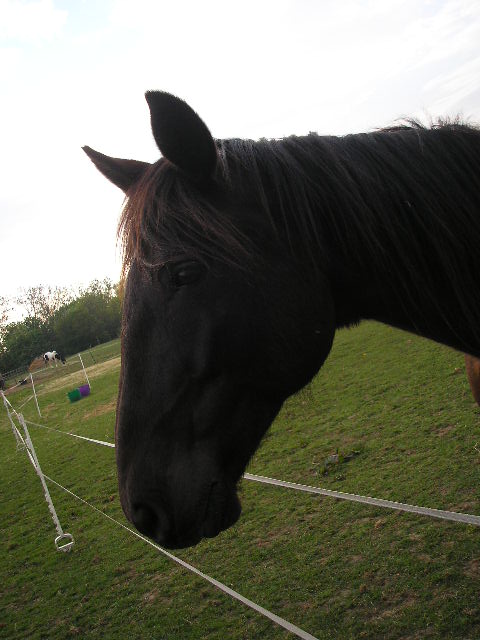
| 08:49, 22 August 2007 | My horse.jpeg (file) |  |
67 KB | Bara | 1 | |
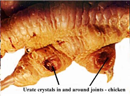
| 19:32, 21 August 2007 | Urate crystals joints.jpg (file) |  |
26 KB | Lizzies | Gross appearance of urate crystals in the joints. Pending permission from Brian Smyth. | 1 |
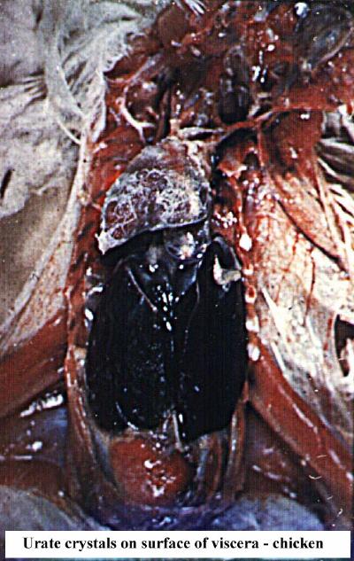
| 19:31, 21 August 2007 | Urate crystals viscera.jpg (file) |  |
59 KB | Lizzies | Gross appearance of urate crystals on the serous membranes. Pending permission from Brian Smyth. | 1 |
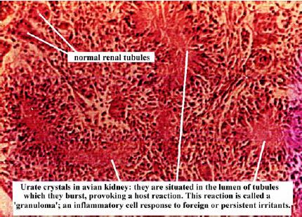
| 19:30, 21 August 2007 | Urate crystals kidney.jpg (file) |  |
50 KB | Lizzies | Histological appearance of urate crystals in the kidney. Pending permission from Brian Smyth. | 1 |
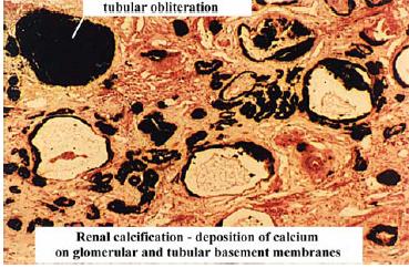
| 19:25, 21 August 2007 | Renal calcification.jpg (file) |  |
34 KB | Lizzies | Histological appearance of calcification in the kidney. Pending permission from Brian Smyth. | 1 |
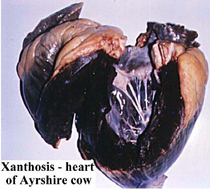
| 18:18, 21 August 2007 | Lipofuscin xanthosis.jpg (file) |  |
30 KB | Lizzies | Xanthosis in the heart of an Ayrshire cow. Pending permission from Brian Smyth. | 1 |
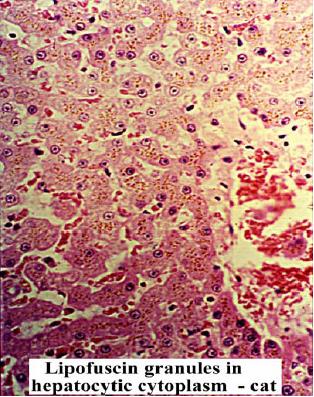
| 18:17, 21 August 2007 | Lipofuscin granules 2.jpg (file) |  |
45 KB | Lizzies | Lipofuscin granules in the cytoplasm of hepatocytes in the cat. Pending permission from Brian Smyth. | 1 |
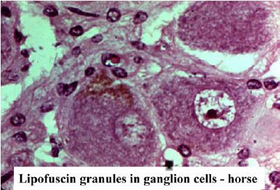
| 18:16, 21 August 2007 | Lipofuscin granules.jpg (file) |  |
28 KB | Lizzies | Histological appearance of lipofuscine granules in ganglion cells of the horse. Pending permission from Brian Smyth. | 1 |
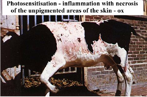
| 18:13, 21 August 2007 | Photosensitisation.jpg (file) |  |
37 KB | Lizzies | Inflammation of the skin due to photosensitisation in the ox. Pending permission from Brian Smyth. | 1 |
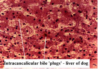
| 18:10, 21 August 2007 | Jaundice bile plugs.jpg (file) |  |
28 KB | Lizzies | Intracanalicular "bile plugs" in the liver of the dog due to jaundice. Pending permission from Brian Smyth. | 1 |
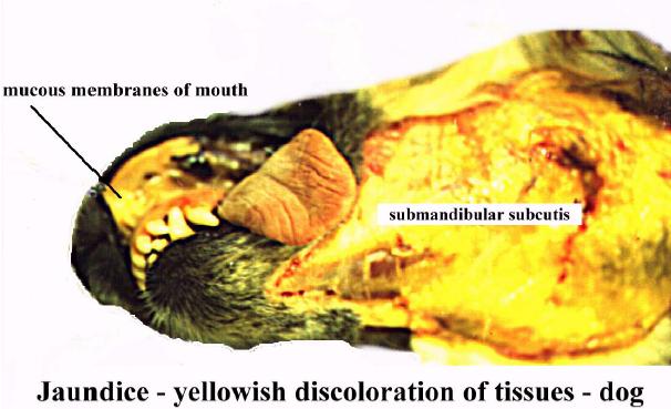
| 18:09, 21 August 2007 | Jaundice discolouration.jpg (file) |  |
38 KB | Lizzies | Yellow discolouration of the mucouse membranse and the mandibular subcutis due to jaundice in the dog. Pending permission from Brian Smyth. | 1 |
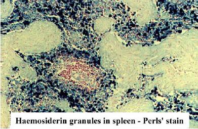
| 17:02, 21 August 2007 | Haemosiderin prussian.jpg (file) |  |
36 KB | Lizzies | Section stained with Prussian Blue to show haemosiderin. Pending permission from Brian Smyth. | 1 |
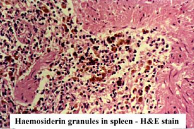
| 14:57, 21 August 2007 | Haemosiderin.jpg (file) |  |
35 KB | Lizzies | Histological appearance of haemosidering granules. Pending permission from Brian Smyth. | 1 |
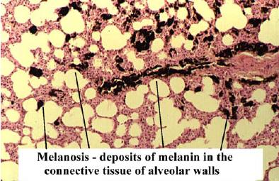
| 13:23, 21 August 2007 | Melanosis histo.jpg (file) |  |
34 KB | Lizzies | Histological appearance of melanosis. Pending permission from Brian Smyth. | 1 |
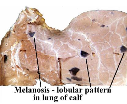
| 13:23, 21 August 2007 | Melanosis gross.jpg (file) |  |
27 KB | Lizzies | Gross appearance of melanosis. Pending permission from Brian Smyth. | 1 |
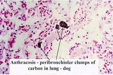
| 12:27, 21 August 2007 | Anthracosis histo.jpg (file) |  |
27 KB | Lizzies | Histological appearance of anthracosis. Pending permission from Brian Smyth. | 1 |
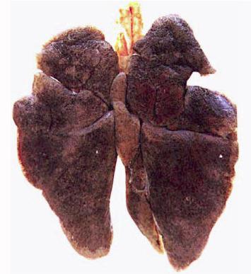
| 12:26, 21 August 2007 | Anthracosis gross.jpg (file) |  |
23 KB | Lizzies | Gross appearance of antrhacosis in the lungs of a dog. Pending permission from Brian Smyth. | 1 |
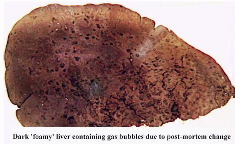
| 12:03, 21 August 2007 | Foamy liver.jpg (file) |  |
27 KB | Lizzies | Appearance of a foamy lover due to putrefaction. Pending permission from Brian Smyth. | 1 |
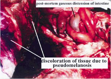
| 12:01, 21 August 2007 | Pseudomelanosis.jpg (file) |  |
24 KB | Lizzies | Appearance of pseudomelanosis. Pending permission from Brian Smyth. | 1 |
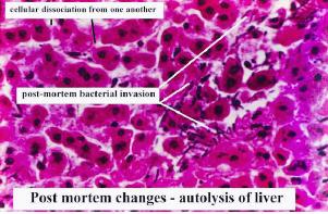
| 10:35, 21 August 2007 | Autolysis of liver.jpg (file) |  |
21 KB | Lizzies | Histological appearance of autolysis in the liver. Pending permission from Brian Smyth. | 1 |
| 09:58, 21 August 2007 | Hypostatic congestion.jpg (file) |  |
11 KB | Lizzies | Hypostatic congestion in the lungs. Pending permission from Brian Smyth. | 1 |
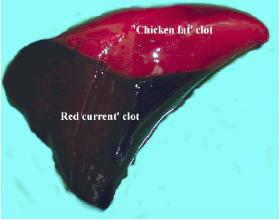
| 09:51, 21 August 2007 | Clot.jpg (file) |  |
8 KB | Lizzies | A post-mortem clot, showing the upper "chicken-fat" clot and the lower "redcurrant-jelly" clot. Pending permission from Brian Smyth. | 1 |
| 06:34, 21 August 2007 | Nick Short.jpg (file) |  |
16 KB | Nshort | 1 | |
| 06:30, 21 August 2007 | Nick Photo.jpg (file) |  |
16 KB | Nshort | Photo of Nick Short | 1 |
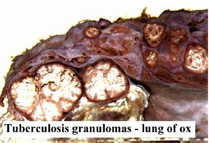
| 21:30, 20 August 2007 | Tuberculosis granuloma.jpg (file) |  |
29 KB | Lizzies | Tuberculosis granuloma. Pending permission from Brian Smyth. | 1 |
| 21:27, 20 August 2007 | Calcification.jpg (file) |  |
19 KB | Lizzies | Histological appearance of calcification. Pending permission from Brian Smyth. | 1 |
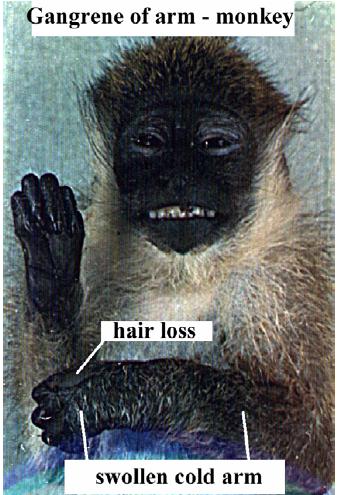
| 21:25, 20 August 2007 | Monkey gangrene 2.jpg (file) |  |
27 KB | Lizzies | Wet gangrene in a monkey (2). Pending permission from Brian Smyth. | 1 |
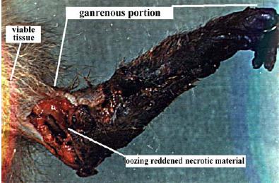
| 21:23, 20 August 2007 | Monkey gangrene 1.jpg (file) |  |
41 KB | Lizzies | Wet gngrene in a monkey (1). Pending permission form Brian Smyth. | 1 |
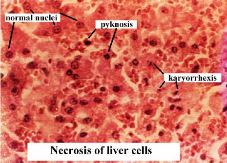
| 21:20, 20 August 2007 | Necrosis histo.jpg (file) |  |
37 KB | Lizzies | Histological appearance of necrosis. Pending permission from Brian Smyth. | 1 |
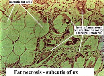
| 21:17, 20 August 2007 | Fat necrosis ox subcutis histo.jpg (file) |  |
33 KB | Lizzies | Histological appearance of fat necrosis in the ox subcutis. Pending permission from Brian Smyth. | 1 |
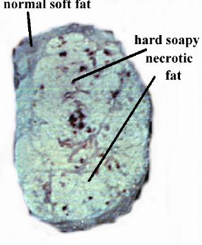
| 21:15, 20 August 2007 | Fat necrosis ox subcutis.jpg (file) |  |
18 KB | Lizzies | Gross appearance of fat necrosis in the ox subcutis. Pending permission from Brian Smyth. | 1 |
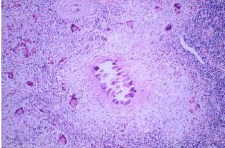
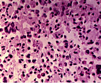
| 21:10, 20 August 2007 | Macrophage caseative necrosis.JPG (file) |  |
39 KB | Lizzies | Histological appearance of caseative necrosis. Note the macrophages. Pending permission from Brian Smyth. | 1 |
| 21:05, 20 August 2007 | Abscess centre and capsule.jpg (file) |  |
19 KB | Lizzies | Gross appearance of an abscess, showing the necrotic centre and the capsule. Pending permission from Brian Smyth. | 1 |
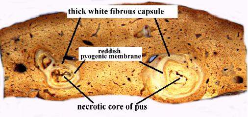
| 21:03, 20 August 2007 | Abscess slice.jpg (file) |  |
31 KB | Lizzies | Slice through and abscess, showing all the layers. Pending permission from Brian Smyth. | 1 |
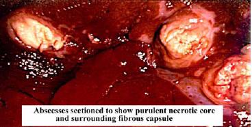
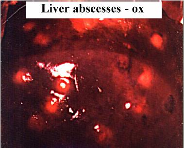
| 21:01, 20 August 2007 | Liver abscess.jpg (file) |  |
21 KB | Lizzies | Abscesses in the liver. Pending permission from Brian Smyth. | 1 |
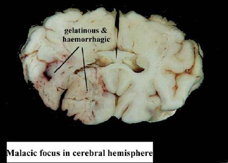
| 20:53, 20 August 2007 | Malacia.jpg (file) |  |
19 KB | Lizzies | 1 | |
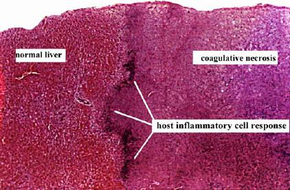
| 20:52, 20 August 2007 | Coagulative necrosis histo.jpg (file) |  |
35 KB | Lizzies | Histological appearance of coagulative necrosis. Pending permission from Brian Smyth. | 1 |
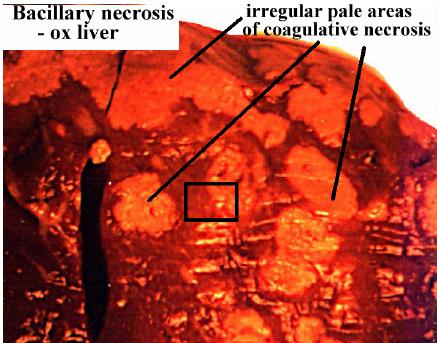
| 20:51, 20 August 2007 | Coagulative necrosis bacillary necrosis.jpg (file) |  |
35 KB | Lizzies | Coagulative necrosis due to bacillary necrosis. Pending permission form Brian Smyth. | 1 |
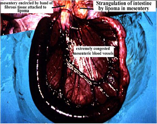
| 20:33, 20 August 2007 | Strangulation of intestine.jpg (file) |  |
49 KB | Lizzies | Stangulation of the intestine by lipoma in the mesentery. Pending permsission from Brian Smyth. | 1 |
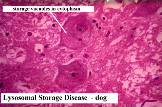
| 19:41, 20 August 2007 | Lysosomal storage disease.jpg (file) |  |
41 KB | Lizzies | Histological appearance of lysomsomal storage disease in the dog. Pending permission from Brian Smyth. | 1 |
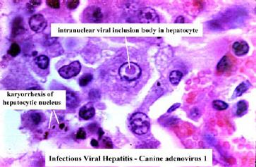
| 19:24, 20 August 2007 | Viral inclusion canine adenovirus 1.jpg (file) |  |
23 KB | Lizzies | Viral inclusion due to canine adenovirus 1. Pending permission from Brain Smyth. | 1 |
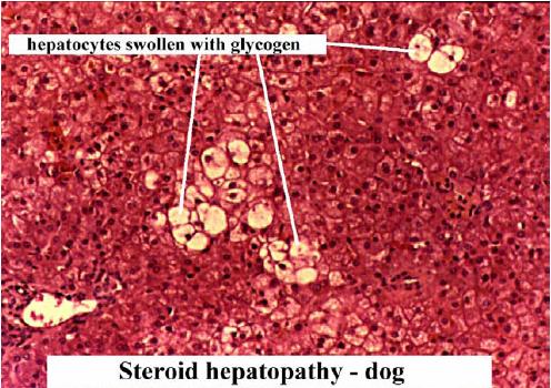
| 19:10, 20 August 2007 | Glycogen infiltration.jpg (file) |  |
53 KB | Lizzies | Glycogen infiltration due to steroid hepatopathy in the dog. Pending permission from Brian Smyth. | 1 |