Unused files
The following files exist but are not embedded in any page. Please note that other web sites may link to a file with a direct URL, and so may still be listed here despite being in active use.
Showing below up to 50 results in range #71 to #120.
View (previous 50 | next 50) (20 | 50 | 100 | 250 | 500)
 Adenoma.jpeg 402 × 652; 83 KB
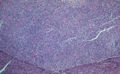
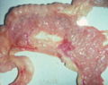
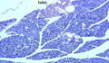
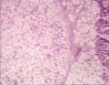
 Perianal gland adenoma histopath.jpg 750 × 576; 100 KB
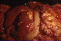
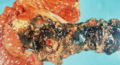
 Perianal gland adenoma.jpg 626 × 570; 51 KB
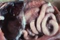
 Strongylus vulgaris.jpg 760 × 548; 78 KB
 Adenoma2.jpeg 652 × 400; 82 KB
 Carcinoma.jpeg 533 × 357; 30 KB
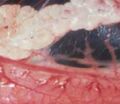
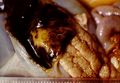
 Carcinoma gross.jpeg 533 × 357; 30 KB
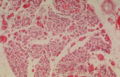
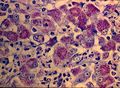
 Carcinoma micro.jpeg 469 × 312; 44 KB
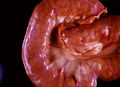
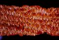
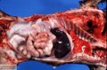
 Infaction of the small bowel.jpg 744 × 576; 65 KB
 Pancreatic nodular hyperplasia.jpeg 510 × 403; 36 KB
 Islet.jpeg 466 × 270; 48 KB
 Pancreatic hypoplasia.jpeg 229 × 199; 10 KB
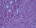
 Pancreatic hypoplasia micro.jpeg 445 × 287; 34 KB
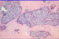
 Brunner gland adenoma.jpg 742 × 574; 75 KB
 Acute haemorrhagic pancreatitis.jpeg 479 × 311; 37 KB
 Acute pancreatic necrosis.jpeg 429 × 234; 30 KB
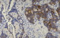
 Insulinoma.jpeg 442 × 287; 56 KB
 Beta cell carcinoma.jpeg 450 × 284; 48 KB
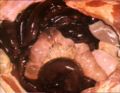
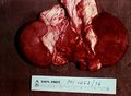
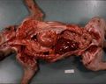
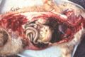
 Atresia ani PM.jpg 807 × 539; 94 KB
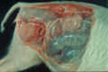
 Gill2.jpg 179 × 202; 14 KB
 Johnes disease proliferative enteritis.jpg 762 × 550; 50 KB
 Pulpy kidney disease.jpg 320 × 256; 15 KB
 Pulpy kidney gross.jpg 762 × 556; 70 KB
 Bucket and spade.jpg 464 × 298; 14 KB
 Trichuris vulpis caecum.jpg 762 × 552; 100 KB
 Trichuris vulpis caecum comparative.jpg 766 × 528; 69 KB
 Trichuris ovis.jpg 762 × 528; 55 KB
 Johnes disease histological.jpg 762 × 558; 100 KB
 Johnes disease proliferative ileitis.jpg 764 × 522; 63 KB
 Porcine intestinal adenomatosis campylobacter.jpg 764 × 528; 107 KB
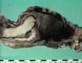
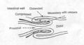 Intussusception.jpg 622 × 332; 34 KB
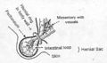 Hernial sac.jpg 713 × 424; 51 KB
 Stomach diaphragmatic hernia.jpg 320 × 256; 13 KB
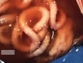
 Volvulus.jpg 762 × 542; 54 KB
 Intussuceptionphoto.jpg 744 × 572; 56 KB
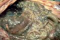
 Chronic peritonitis with fibrosis.jpeg 750 × 491; 74 KB
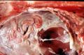
 FIP.jpeg 750 × 500; 55 KB
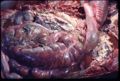
 Omentum carcinoma.jpeg 750 × 504; 308 KB
 Bile stained peritonitis and gastric rupture.jpeg 750 × 497; 66 KB
 FIP severe exudative peritonitis.jpeg 750 × 571; 36 KB
 Nocardiosis in a puma.jpeg 750 × 497; 310 KB
 Glasser's disease - severe acute fibrinous peritonitis.jpeg 750 × 493; 75 KB
 Acute peritonitis and cecal base rupture.jpeg 750 × 508; 100 KB
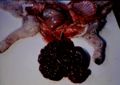
 Cysticercus pisiformis.jpeg 750 × 497; 276 KB
 Tubeculous peritonitis.jpeg 320 × 248; 13 KB
 Carcinomatosis and sclerosis in sheep.jpeg 750 × 499; 189 KB
 Lipoma in horse.jpeg 750 × 499; 63 KB
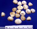 Bovine pancreatic calculi.jpeg 750 × 594; 50 KB
 Pancreatic hypoplasia by King.jpeg 750 × 513; 50 KB


