Unused files
The following files exist but are not embedded in any page. Please note that other web sites may link to a file with a direct URL, and so may still be listed here despite being in active use.
Showing below up to 50 results in range #351 to #400.
View (previous 50 | next 50) (20 | 50 | 100 | 250 | 500)
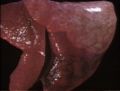
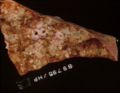
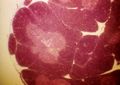
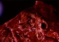
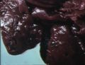
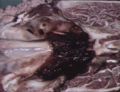
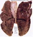 Melanoma metastases dog lung.jpg 426 × 464; 44 KB
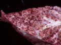
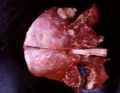
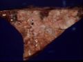
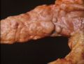
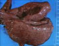
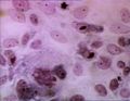
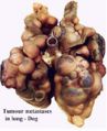 Tumour metastases lung dog.jpg 382 × 470; 30 KB
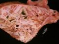
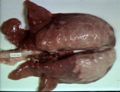
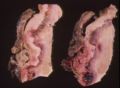
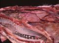
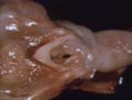
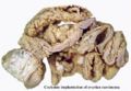 Coelomic implantation ovarian carcinoma.jpg 549 × 380; 36 KB
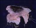
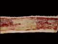
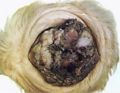 Squamous cell carcinoma eye.jpg 394 × 304; 23 KB
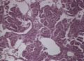
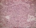
 Squamous cell carcinoma histo.jpg 552 × 357; 61 KB
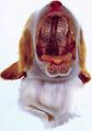
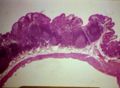
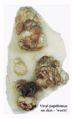 Viral papilloma.jpg 358 × 593; 27 KB
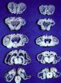 Swayback sections.jpg 361 × 494; 40 KB
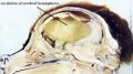
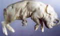 Cyclops.jpg 554 × 333; 35 KB
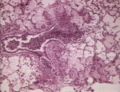 Acute exudative pneumonia.jpg 320 × 246; 21 KB
 Acute exudative pneumonia - gross.jpg 320 × 242; 11 KB
 Acute fibrinous pneumonia.jpg 320 × 240; 14 KB
 Cleft palate.jpg 328 × 465; 21 KB
 Hydrocephalus.jpg 809 × 452; 67 KB
 Acute necrotising pneumonia.jpg 732 × 570; 55 KB
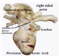 Persistent right aortic arch.jpg 471 × 447; 32 KB
 Acute suppurative pneumonia.jpg 742 × 574; 50 KB
 Adenomatosis of lung.jpg 744 × 544; 67 KB
 Adenovirus pneumonia.jpg 746 × 570; 50 KB
 Alveolar cell carcinoma.jpg 744 × 572; 83 KB
 Alveolar emphysema.jpg 758 × 574; 83 KB
 Aspergillus pneumonia of cattle.jpg 744 × 574; 63 KB
 Bronchiectasis.jpg 320 × 246; 20 KB
 Bronchiectasis micro.jpg 748 × 574; 103 KB
 Bronchiolitis obliterans.jpg 750 × 558; 63 KB
 Chronic bronchiolitis.jpg 746 × 572; 60 KB
 Thymus histo.jpg 758 × 540; 68 KB
 Peyers patches.jpg 758 × 556; 57 KB
 Chronic bronchopneumonia.jpg 750 × 564; 47 KB
 Calf pneumonia.jpg 746 × 554; 60 KB
 Collapsed trachea.jpg 742 × 576; 50 KB
 Contagious bovine pleuropneumonia.jpg 762 × 548; 48 KB
 Destructive emphysema - horse.jpg 752 × 574; 54 KB
 Enzootic pneumonia of pigs.jpg 742 × 570; 57 KB
 Fibrosarcoma in rhinarium of cat.jpg 744 × 576; 48 KB
 Gangrenous pneumonia.jpg 750 × 574; 59 KB
 Granulomatous mycotic pneumonia.jpg 746 × 576; 62 KB
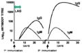 Primary and secondary response.jpg 561 × 362; 25 KB
 Guttural pouch mycosis.jpg 742 × 544; 54 KB
 Haemorrhage in GP mycosis.jpg 746 × 570; 68 KB
 Inclusion body rhinitis.jpg 744 × 576; 57 KB
 IBR nasal cavity.jpg 746 × 548; 70 KB
 IBR trachea.jpg 748 × 572; 38 KB
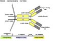 Antibody structure.jpg 658 × 438; 34 KB
 Inhalation pneumonia.jpg 742 × 574; 89 KB
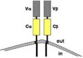 TcR structure.jpg 399 × 279; 11 KB
 Interstitial pneumonia micro.jpg 748 × 480; 49 KB
 Laryngeal oedema.jpg 744 × 564; 49 KB
 Lipid pneumonia.jpg 744 × 570; 74 KB
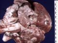 Lung carcinoma.jpg 742 × 552; 70 KB
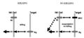 NK cell killing.jpg 604 × 270; 16 KB


