Uploads by Ctrace
Jump to navigation
Jump to search
This special page shows all uploaded files.
| Date | Name | Thumbnail | Size | Description | Versions |
|---|---|---|---|---|---|
| 11:26, 3 June 2011 | GIT-1.swf (file) | 1.97 MB | {{Information |Description=PowerPoint tutorial on Gastrointestinal tract histology, converted to swf format. Resource page can be found here |Source=The Royal Veterinary College |Date=2011 |Author=[[R | 1 | |
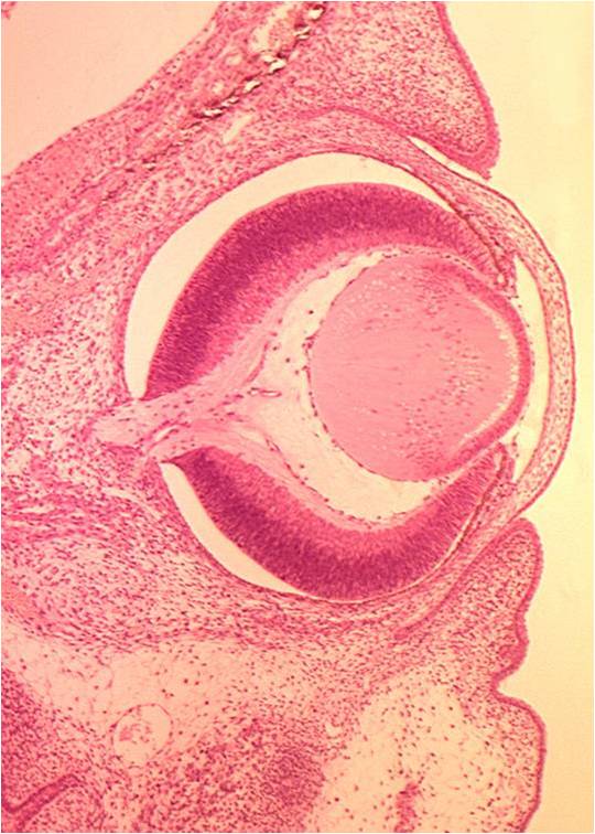
| 10:04, 3 June 2011 | Eye and ear.jpg (file) |  |
76 KB | {{Information |Description=Screenshot from Powerpoint, courtesy of the Royal Veterinary College. Shows section through developing eye. |Source=The Royal Veterinary College |Date=03/06/2011 |Author=Chris | 1 |
| 10:01, 3 June 2011 | Eye and Ear.swf (file) | 1.92 MB | == Summary == {{Information |Description=PowerPoint tutorial on eye and ear histology, converted to swf format. Resource page can be found here |Source=The Royal Veterinary College |Date=2011 |Author=[[RVC|The R | 1 | |
| 10:23, 31 May 2011 | Mair Cover.jpg (file) |  |
15 KB | {{Information |Description =Screen shot of the Equine Internal Medicine SACR book from Manson Publishing. Used as part of the OVAL Project |Source =[http://www.mansonpublishing.com/vet_titles/Mair.html Manson Publishing] |Author =M | 1 |
| 08:38, 31 May 2011 | Follicle.jpg (file) |  |
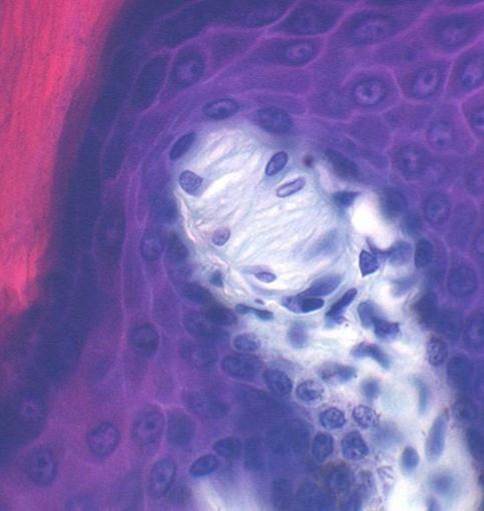
44 KB | {{Information |Description=Screenshot from Powerpoint, courtesy of the Royal Veterinary College. Image shows hair follicle |Source=The Royal Veterinary College |Date=31/05/2011 |Author=Chris Trace |Permis | 1 |
| 08:33, 31 May 2011 | Integument 2.swf (file) | 1.91 MB | {{Information |Description=PowerPoint tutorial on Integument Histology, converted to swf format. Resource page can be found here |Source=The Royal Veterinary College |Date=2011 |Author=John Bredl |Permission=See | 1 | |
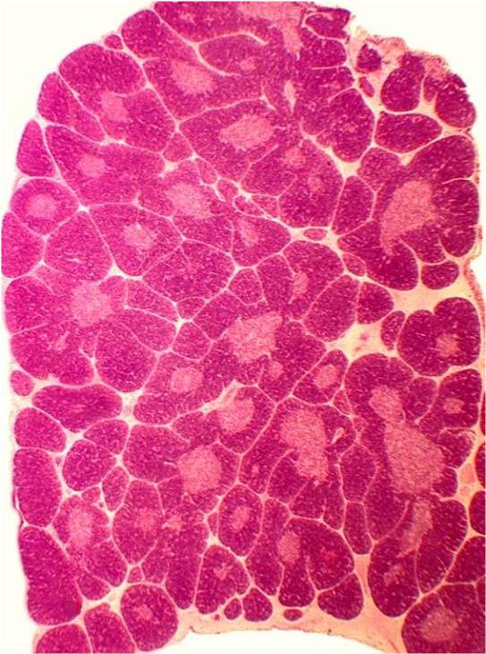
| 19:34, 30 May 2011 | Thymus.jpg (file) |  |
82 KB | {{Information |Description=Screenshot from Powerpoint, courtesy of the Royal Veterinary College. Shows histological section of the thymus from a cat. |Source=The Royal Veterinary College |Date=30/05/2011 | 1 |
| 19:29, 30 May 2011 | Lymphoid Tissue.swf (file) | 1.89 MB | {{Information |Description=PowerPoint tutorial on lymphoid tissue histology, converted to swf format. Resource page can be found here |Source=The Royal Veterinary College |Date=2011 |Author=John Bredl |Permi | 1 | |
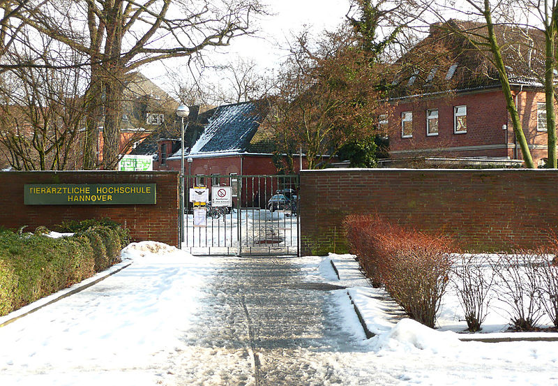
| 10:41, 28 May 2011 | Tiho Eingang.jpg (file) |  |
189 KB | {{Information |Description={{de|Eingang Tierärztliche Hochschule Hannover am Braunschweiger Platz<br/> }} |Source=Transferred from [http://de.wikipedia.org de.wikipedia]; transferred to Commons by User:Ireas using [http://tools.wikimedia.de/~magnus/c | 1 |
| 10:21, 28 May 2011 | TiHo3.jpg (file) |  |
629 KB | {{Information |Description =Logo for Stiftung Tierärztliche Hochschule Hannover |Source =Stiftung Tierärztliche Hochschule Hannover - found on [http://commons.wikimedia.org/wiki/File:TIHO_LogoStiftung_.jpg WikiMedia Commons] |Author = | 1 |
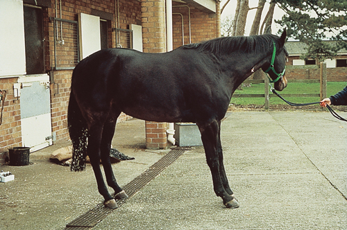
| 15:45, 24 May 2011 | Equine Orthopaedics and Rheumatology 01b.jpg (file) |  |
322 KB | {{Information |Description=Image from [http://www.mansonpublishing.com/vet_titles/May.html 'Equine Orthopaedics and Rheumatology'], with permission from Manson Publishing, as part of the OVAL Project. This is an image of a discharge from t | 1 |
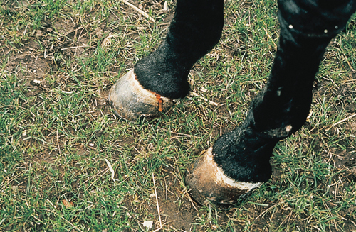
| 15:40, 24 May 2011 | Equine Orthopaedics and Rheumatology Q&A 01a.jpg (file) |  |
240 KB | {{Information |Description=Image from [http://www.mansonpublishing.com/vet_titles/May.html 'Equine Orthopaedics and Rheumatology'], with permission from Manson Publishing, as part of the OVAL Project. This is an image of a severely laminit | 1 |
| 15:27, 24 May 2011 | May Cover.jpg (file) |  |
15 KB | {{Information |Description =Screen shot of the Equine Orthopaedics and Rheumatology SACR book from Manson Publishing. Used as part of the OVAL Project |Source =[http://www.mansonpublishing.com/vet_titles/May.html Manson Publishing] |Author | 1 |
| 13:33, 24 May 2011 | Muscle Tissue.swf (file) | 2 MB | {{Information |Description =PowerPoint tutorial on Muscle Tissue Histology, converted to swf format. Resource page can be found here |Source =The Royal Veterinary College |Author =John Bred | 1 | |
| 10:53, 24 May 2011 | NERVE CELLS.swf (file) | 1.99 MB | {{Information |Description =PowerPoint tutorial on Nerve Cell Histology, converted to swf format. Resource page can be found here |Source =The Royal Veterinary College |Author =John Bredl |Da | 1 | |
| 09:28, 24 May 2011 | Blood and Haemopoiesis.swf (file) | 1.9 MB | {{Information |Description =Blood and Haemopoiesis powerpoint tutorial, converted into swf format |Source =Royal Veterinary College |Author =John Bredl |Date =2011 |Permission = | 1 | |
| 08:49, 24 May 2011 | Connective Tissue.swf (file) | 1.99 MB | {{Information |Description =PowerPoint tutorial on Connective Tissue Histology, converted to swf format. Resource page can be found here |Source =The Royal Veterinary College |Author =Jo | 1 | |
| 11:23, 23 May 2011 | Epithelia Histology.swf (file) | 1.97 MB | {{Information |Description =Epithelial histology powerpoint tutorial converted into flash, this new version has incorporated navigation |Source =Royal Veterinary College |Author =John Bredl |Date =23/05/2011 |Permissi | 1 | |
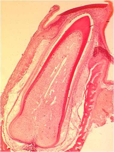
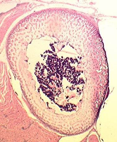
| 13:46, 22 May 2011 | Oral Cavity 2.jpg (file) |  |
35 KB | {{Information |Description=Screenshot from Powerpoint, courtesy of the Royal Veterinary College. Shows longitudinal section through developing tooth. |Source=The Royal Veterinary College |Date=22/05/2011 | 1 |
| 13:41, 22 May 2011 | Oral Cavity 2.swf (file) | 1.96 MB | {{Information |Description=PowerPoint tutorial on oral cavity histology, converted to swf format. Resource page can be found here |Source=The Royal Veterinary College |Date=2011 |Author=[[RVC|The Royal Veterinar | 1 | |
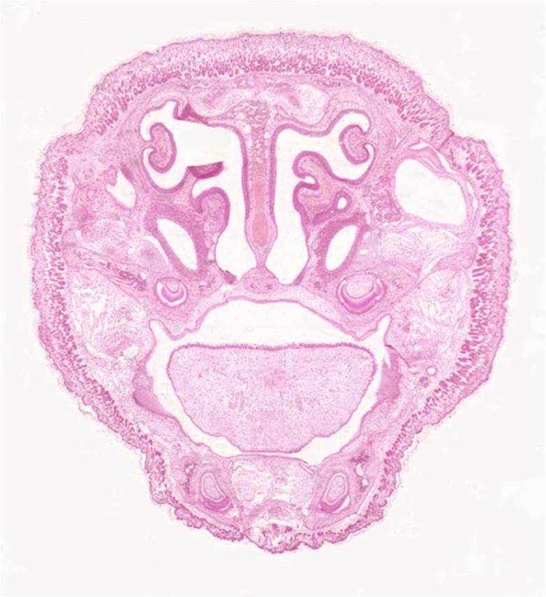
| 11:35, 22 May 2011 | Oral cavity.jpg (file) |  |
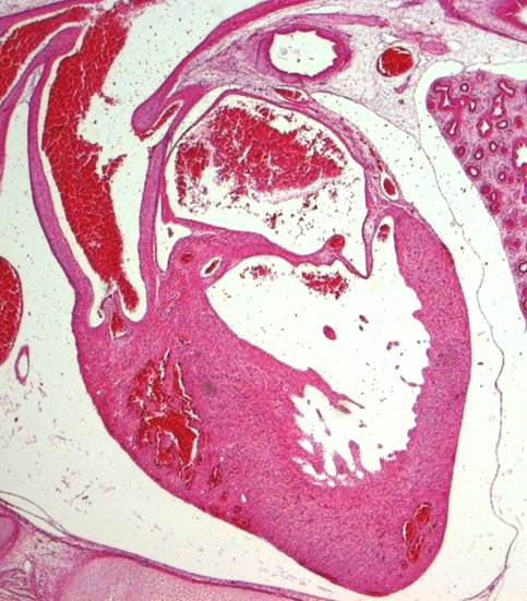
69 KB | {{Information |Description=Screenshot from Powerpoint, courtesy of the Royal Veterinary College. Shows transverse section through embryonic head at level of nasal cavity |Source=[[RVC|The Royal Veterinary Colleg | 1 |
| 11:28, 22 May 2011 | Oral Cavity 1.swf (file) | 1.96 MB | {{Information |Description=PowerPoint tutorial on oral cavity histology, converted to swf format. Resource page can be found here |Source=The Royal Veterinary College |Date=2011 |Author=[[RVC|The Royal Veterinar | 1 | |
| 14:43, 19 May 2011 | Dragster jigsaw.jpg (file) |  |
17 KB | {{Information |Description=Blue jigsaw piece, dragster logo |Source=RVC |Date=19/05/2011 |Author=Chris Trace |Permission=See below |Other_versions= }} | 1 |
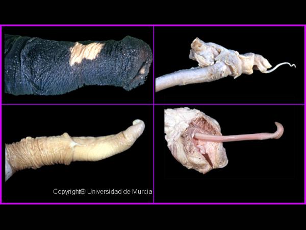
| 12:26, 19 May 2011 | Penises.jpg (file) |  |
31 KB | {{Information |Description =Image of a an equine, feline, bovine and ovine penises. |Source =Murcia Vet School |Author =[[Spain - Universidad de Murcia Facultad de Veterinaria|Mu | 1 |
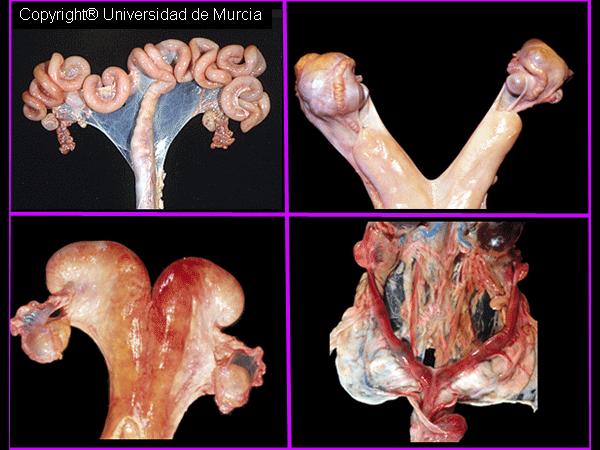
| 12:14, 19 May 2011 | Uteri.jpg (file) |  |
47 KB | {{Information |Description =Image of a an equine, feline, bovine and ovine uteri. |Source =Murcia Vet School |Author =[[Spain - Universidad de Murcia Facultad de Veterinaria|Murc | 1 |
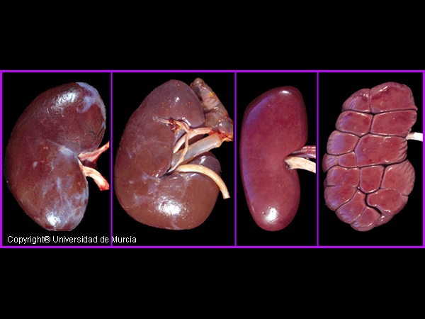
| 12:02, 19 May 2011 | Kidneys.jpg (file) |  |
96 KB | {{Information |Description =Image of a an equine, canine, ruminant and porcine kidney. |Source =Murcia Vet School |Author =[[Spain - Universidad de Murcia Facultad de Veterinaria | 1 |
| 11:51, 19 May 2011 | Stomachs.jpg (file) |  |
31 KB | {{Information |Description =Image of a an equine, canine and porcine stomach. |Source =Murcia Vet School |Author =[[Spain - Universidad de Murcia Facultad de Veterinaria|Murcia V | 1 |
| 11:34, 19 May 2011 | ComparativeLivers.jpg (file) |  |
37 KB | {{Information |Description =Image of a ruminant, equine, canine and porcine liver. |Source =Murcia Vet School |Author =[[Spain - Universidad de Murcia Facultad de Veterinaria|Mur | 1 |
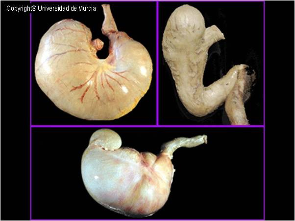
| 11:23, 19 May 2011 | Comparative stomachs.jpg (file) |  |
42 KB | {{Information |Description =Image of a ruminant abomasum and an equine, canine and porcine stomach. |Source =Murcia Vet School |Author =[[Spain - Universidad de Murcia Facultad d | 1 |
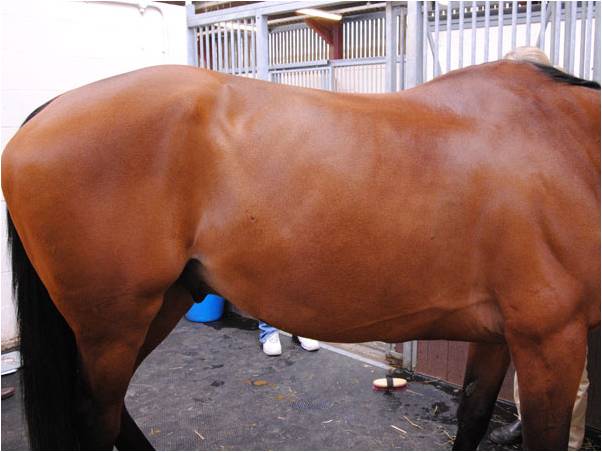
| 11:08, 19 May 2011 | EquineAbdomen.jpg (file) |  |
35 KB | {{Information |Description =Image of a bay horse's right abominal flank |Source =Royal Veterinary College |Author =Royal Veterinary College |Date =2010 |Permission = |other_versions = }} | 1 |
| 10:36, 19 May 2011 | Dragster2.jpg (file) |  |
19 KB | {{Information |Description=screenshot of dragster activity |Source=http://www.rvc.ac.uk/Review/Dragster/index.html |Date=08/12/10 |Author=RVC |Permission=See below |Other_versions= }} | 1 |
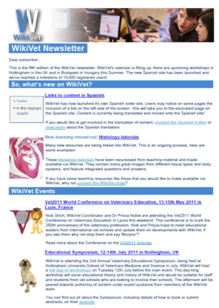
| 09:39, 19 May 2011 | Issue5.jpg (file) |  |
45 KB | {{Information |Description =screenshot of WikiVet newsletter Issue 5 |Source =WikiVet newsletter Issue 5 |Author =Chris Trace |Date =newsletter sent 12th May 2011 |Permission = |other_versions = }} | 1 |
| 09:32, 19 May 2011 | WikiVet Newsletter Issue 5.pdf (file) | 142 KB | {{Information |Description =5th Issue of the WikiVet newsletter |Source =WikiVet |Author =Chris Trace |Date =Sent 12th May 2011 |Permission = |other_versions = }} | 1 | |
| 09:34, 12 May 2011 | Workshop.png (file) |  |
137 KB | {{Information |Description =Logo for WikiVet workshops |Source =Royal Veterinary College |Author =Chris Trace |Date =11/05/2011 |Permission = |other_versions = }} | 1 |
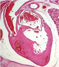
| 10:07, 11 May 2011 | Heart.jpg (file) |  |
14 KB | {{Information |Description=Screenshot from Powerpoint, courtesy of the Royal Veterinary College. Shows a longitudinal histological section of a heart |Source=The Royal Veterinary College |Date= | 1 |
| 12:26, 9 May 2011 | Bone and Cartilage2.jpg (file) |  |
38 KB | {{Information |Description=Screenshot from secondBone and Cartilage Powerpoint tutorial, courtesy of the Royal Veterinary College. Image shows transverse section of diaphysis of developing rabbit bone |So | 1 |
| 12:19, 9 May 2011 | Bone and Cartilage2.swf (file) | 1.88 MB | {{Information |Description=2nd PowerPoint tutorial on Bone and Cartilage Histology, converted to swf format. Resource page can be found here |Source=The Royal Veterinary College |Date=2011 |Author=[[RVC|T | 1 | |
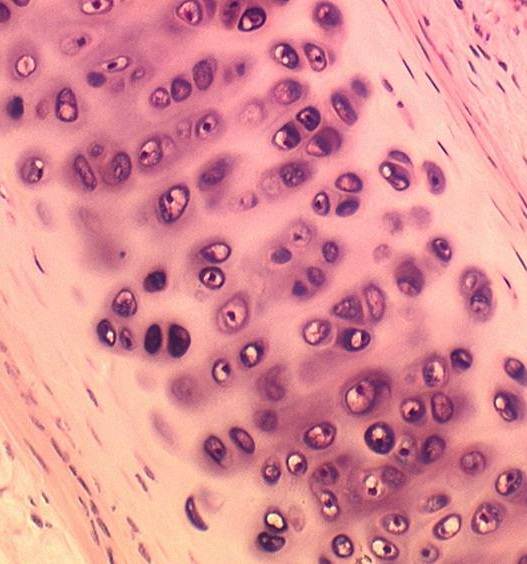
| 11:28, 9 May 2011 | Bone and Cartilage1.jpg (file) |  |
42 KB | {{Information |Description=Screenshot from firstBone and Cartilage Powerpoint tutorial, courtesy of the Royal Veterinary College. Image shows hyaline cartilage from a trachea |Source=[[RVC|The Royal Veter | 1 |
| 11:20, 9 May 2011 | Bone and Cartilage.swf (file) | 1.73 MB | {{Information |Description=PowerPoint tutorial on Bone and Cartilage Histology, converted to swf format. Resource page can be found here |Source=The Royal Veterinary College |Date=2011 |Author=[[RVC|The R | 1 | |
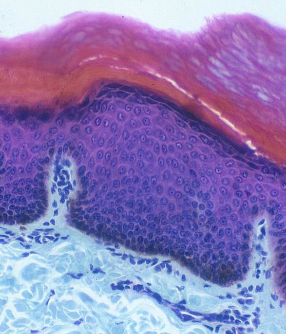
| 08:50, 9 May 2011 | Integument.jpg (file) |  |
57 KB | {{Information |Description=Screenshot from Powerpoint, courtesy of the Royal Veterinary College. Image shows layers of epidermis |Source=The Royal Veterinary College |Date=09/05/2011 |Author=Chris Trace | | 1 |
| 08:41, 9 May 2011 | Integument.swf (file) | 1.84 MB | {{Information |Description=PowerPoint tutorial on Integument Histology, converted to swf format. Resource page can be found here |Source=The Royal Veterinary College |Date=2011 |Author=[[RVC|The Royal Veterinary | 1 | |
| 16:48, 8 May 2011 | Cardiovascular System.jpg (file) |  |
57 KB | {{Information |Description=Screenshot from Powerpoint, courtesy of the Royal Veterinary College. Shows a longitudinal histological section of a heart |Source=The Royal Veterinary College |Date= | 1 |
| 16:41, 8 May 2011 | Cardiovascular system.swf (file) | 2 MB | {{Information |Description=PowerPoint tutorial on cardiovascular system histology, converted to swf format. Resource page can be found here |Source=The Royal Veterinary College |Date=2011 |Author=[[RVC | 1 | |
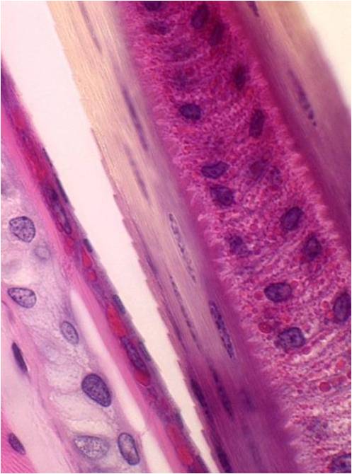
| 11:53, 8 May 2011 | Respiratory system.jpg (file) |  |
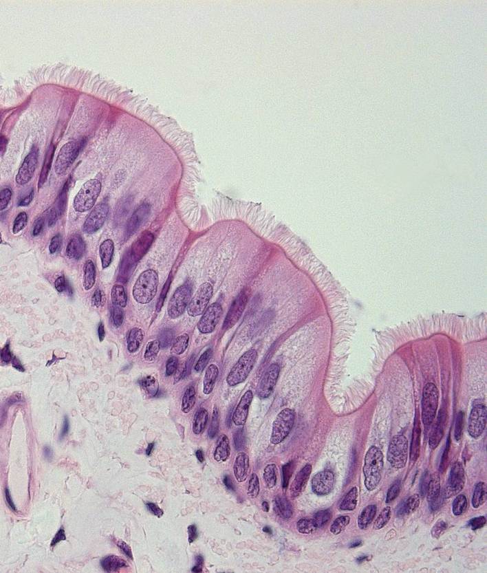
87 KB | {{Information |Description=Screenshot from Powerpoint, courtesy of the Royal Veterinary College. Shows cilia and pseudostratified epithelium |Source=The Royal Veterinary College |Date=08/05/2011 | | 1 |
| 11:51, 8 May 2011 | Respiratory system.swf (file) | 1.86 MB | {{Information |Description=PowerPoint tutorial on respiratory system histology, converted to swf format. Resource page can be found here |Source=The Royal Veterinary College |Date=2011 |Author=[[RVC|The R | 1 | |
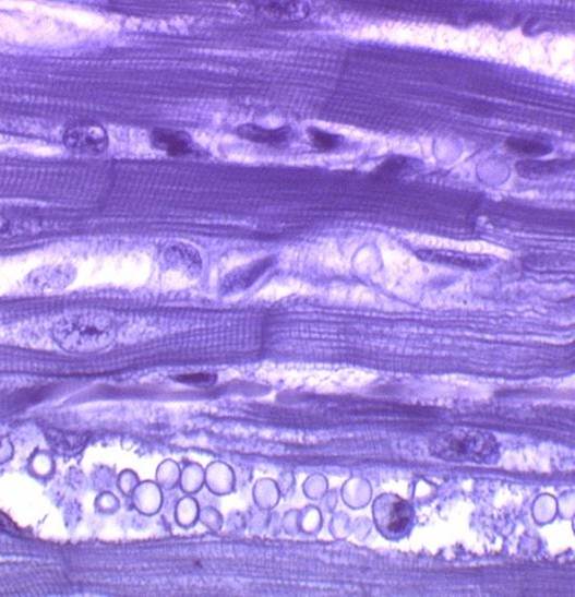
| 11:09, 8 May 2011 | Muscle tissue.jpg (file) |  |
44 KB | {{Information |Description=Screenshot from Powerpoint, courtesy of the Royal Veterinary College |Source=The Royal Veterinary College |Date=08/05/2011 |Author=Chris Trace |Permission=See below |Other_ver | 1 |
| 10:06, 8 May 2011 | Nerve cells.jpg (file) |  |
26 KB | {{Information |Description=Screenshot from Powerpoint, courtesy of the Royal Veterinary College |Source=The Royal Veterinary College |Date=08/05/2011 |Author=Chris Trace |Permission=See below |Other_versi | 1 |
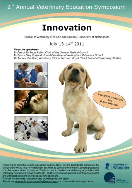
| 12:50, 6 May 2011 | Nottingham poster.jpg (file) |  |
45 KB | {{Information |Description=Image of poster for the 2nd Annual Veterinary Education Symposium |Source=Nottingham Vet school |Date=2011 |Aut | 1 |
| 12:44, 6 May 2011 | Nottingham poster.pdf (file) | 515 KB | {{Information |Description=Poster for the 2nd Annual Veterinary Education Symposium |Source=Nottingham Vet school |Date=2011 |Author=Liz M | 1 | |
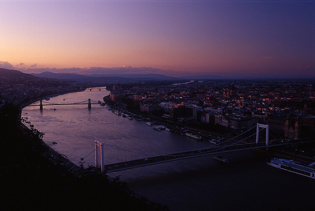
| 13:18, 4 May 2011 | Budapest.jpg (file) |  |
65 KB | {{Information |Description =Photo of Budapest |Source =[http://www.flickr.com/photos/9702937@N06/1418473231/ Flickr] |Author =mck...'s |Date =4th August 2007 |Permission = |other_versions = }} Licensed under a [http://crea | 1 |