Uploads by Lizzies
Jump to navigation
Jump to search


This special page shows all uploaded files.


| Date | Name | Thumbnail | Size | Description | Versions |
|---|---|---|---|---|---|
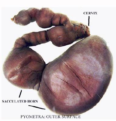
| 10:09, 31 August 2007 | Pyometra.jpg (file) |  |
23 KB | Pyometra - outside surface of the uterus. Pending permission from Brian Smyth. | 1 |
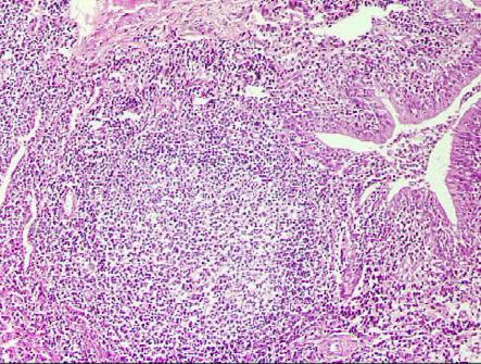
| 09:54, 31 August 2007 | Lymphoid follicle lung.jpg (file) |  |
64 KB | Lymphoid follicle in the lung. Has developed following extensive lymphocytic infiltration. Pending permission from Brian Smyth. | 1 |
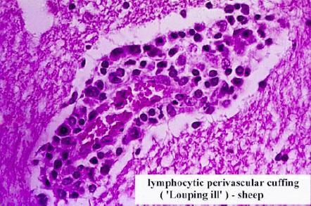
| 09:51, 31 August 2007 | Perivascular cuffing.jpg (file) |  |
44 KB | Lymphocytic perivascular cuffing in louping ill in the sheep. Pending permission from Brian Smyth. | 1 |
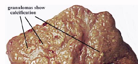
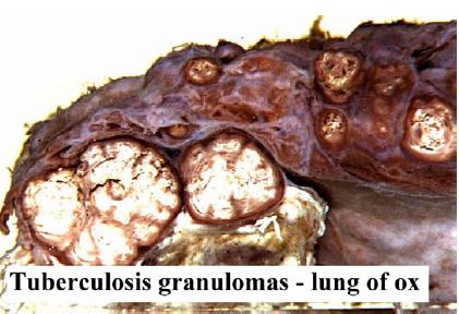
| 09:48, 31 August 2007 | Tuberculous granulomas.jpg (file) |  |
20 KB | Gross appearance of tuberculous granulomas in the lymph node of the ox. These granulomas show a degree of calcification. Pending permission fromo Brian Smyth. | 1 |
| 09:45, 31 August 2007 | Lumpy jaw histology.jpg (file) |  |
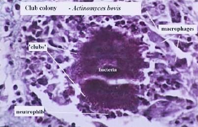
26 KB | Structure of a granuloma in Lumpy Jaw (actinomycosis). Note the central core of bacteria, surrounded by "clubs", followed by a layer of inflammatory cells (neutrophils and macrophages). Pending permission from Brian Smyth. | 1 |
| 09:43, 31 August 2007 | Lumpy jaw mandible.jpg (file) |  |
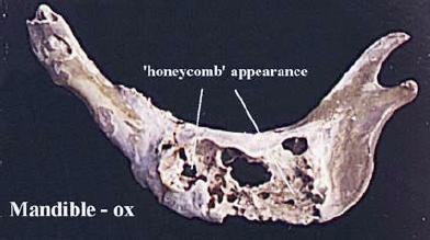
17 KB | "Honeycomb" appearance of mandible, following actinomycosis (Lumpy Jaw). Pending permission from Brian Smyth. | 1 |
| 09:42, 31 August 2007 | Lumpy jaw presentation.jpg (file) |  |
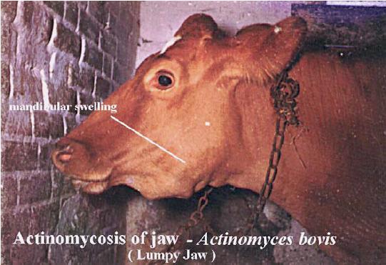
41 KB | Cow showing actinomycosis of the jaw (Lumpy Jaw). Note the mandibular swelling. Pending permission from Brian Smyth. | 1 |
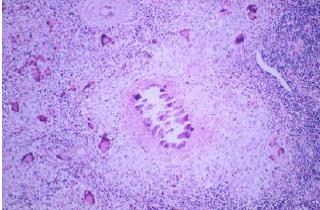
| 09:37, 31 August 2007 | Langhans giant cell.jpg (file) |  |
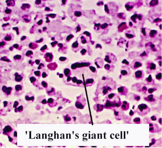
54 KB | Langhans giant cell. Pending permission from Brian Smyth. | 1 |
| 09:34, 31 August 2007 | Macrophages.jpg (file) |  |
15 KB | Macrophages. Pending permission from Brian Smyth. | 1 |
| 09:32, 31 August 2007 | Lymphocytes and plasma cell.jpg (file) |  |
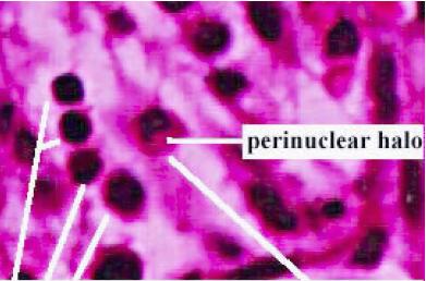
21 KB | Note the 4 lymphocytes on the right hand side, and the plasma cell with the perinuclear halo. Pending permission from Brian Smyth. | 1 |
| 09:29, 31 August 2007 | Mast cells.jpg (file) |  |
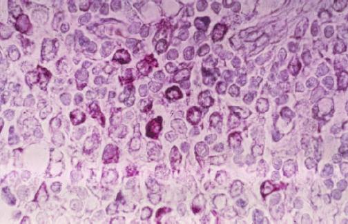
37 KB | Mast cells. Pending permission from Brian Smyth. | 1 |
| 09:28, 31 August 2007 | Eosinophils.jpg (file) |  |
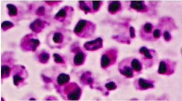
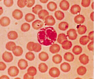
17 KB | Eosinophils (H&E). Pending permission from Brian Smyth. | 1 |
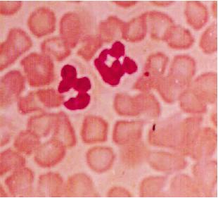
| 09:28, 31 August 2007 | Neutrophils.jpg (file) |  |
13 KB | Neutrophils (H&E). Pending permission from Brian Smyth. | 1 |
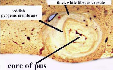
| 09:23, 31 August 2007 | Acute abscess.jpg (file) |  |
23 KB | Cross section through an acute abscess. Pending permission from Brian Smyth. | 1 |
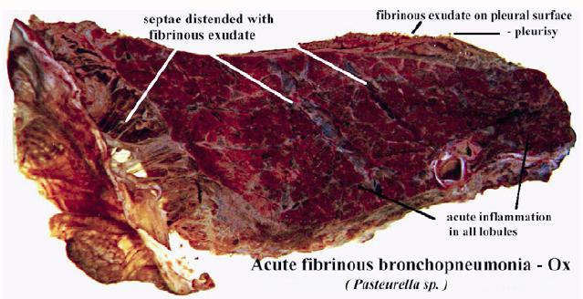
| 09:20, 31 August 2007 | Acute fibrinous bronchopneumonia.jpg (file) |  |
47 KB | Gross appearance on acute fibrinous bronchopneumonia in the ox, caused by ''Pasteurella sp.''. Note the septae are distended with fibrinous exudate. Pending permission from Brian Smyth. | 1 |
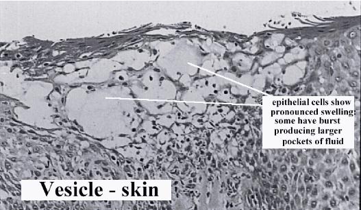
| 09:14, 31 August 2007 | Serous fluid vesicle.jpg (file) |  |
43 KB | Histological appearance of a vesicle in the skin, containting serous exudate. Pending permission from Brian Smyth. | 1 |
| 16:34, 29 August 2007 | Haemorrhage in the brain.jpg (file) |  |
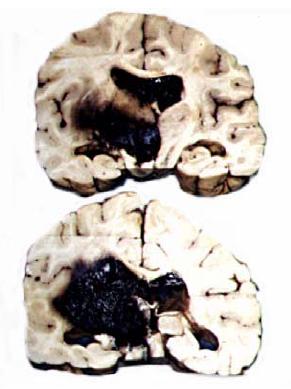
19 KB | Haemorrhage in the brain. Pending permission from Brian Smyth. | 1 |
| 16:30, 29 August 2007 | Anasarca.jpg (file) |  |
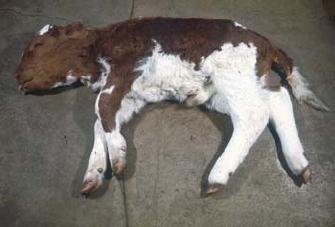
12 KB | Anasarca. Pending permission from Brian Smyth. | 1 |
| 16:20, 29 August 2007 | Pulmonary oedema.jpg (file) |  |
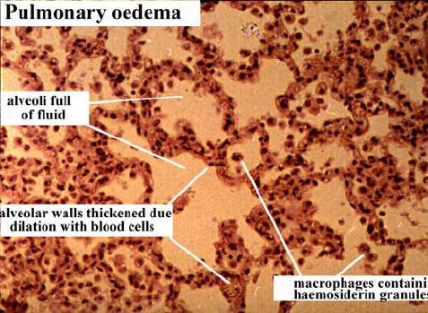
48 KB | Histological appearance of pulmonary oedema. Pending permission from Brian Smyth. | 1 |
| 12:23, 29 August 2007 | Thrombosis strongylus vulgaris.jpg (file) |  |
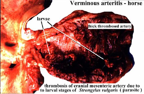
49 KB | Thrombosis in the cranial mesenteric artery, caused by ''Strongylus vulgaris'' larvae. Pending permission from Brian Smyth. | 1 |
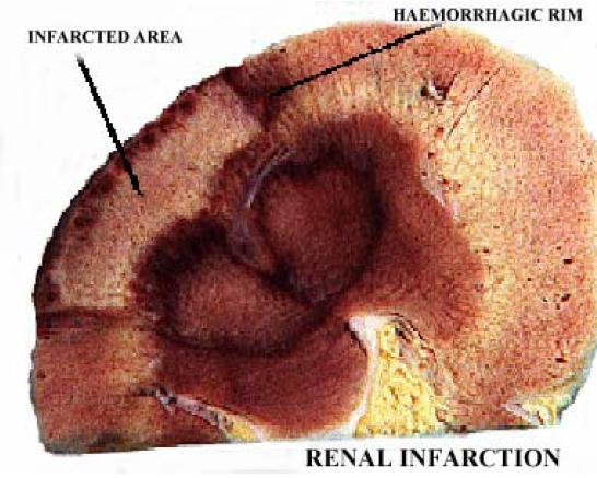
| 12:21, 29 August 2007 | Renal infarction.jpg (file) |  |
46 KB | Gross appearance of renal infarction. Pending permission from Brian Smyth. | 1 |
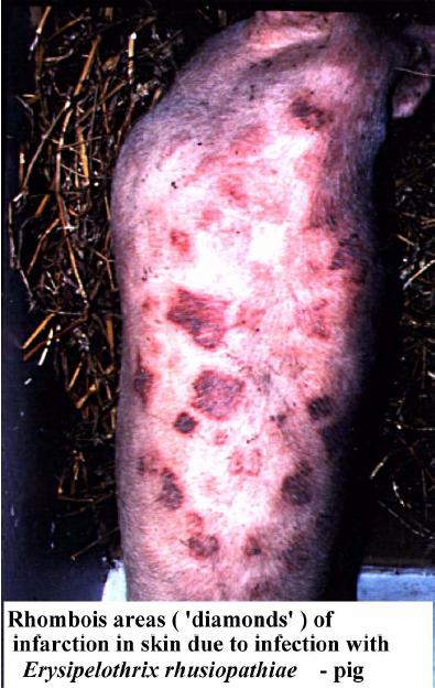
| 12:20, 29 August 2007 | Skin infarction pig.jpg (file) |  |
51 KB | Infarction in the skin of the pig caused by ''Erysipelothrix rhusiopathiae''. Pending permission from Brian Smyth. | 1 |
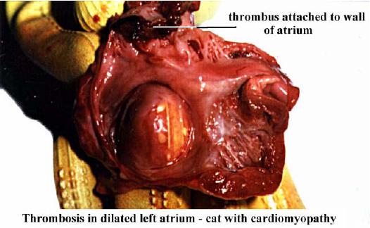
| 12:09, 29 August 2007 | Thrombus cat left atrium.jpg (file) |  |
31 KB | Gross appearance of a thrombus attached to the wall of the left atrium in a cat suffering from cardiomyopathy. Pending permission from Brian Smyth. | 1 |
| 09:23, 22 August 2007 | Cocktails.jpg (file) |  |
30 KB | 1 | |
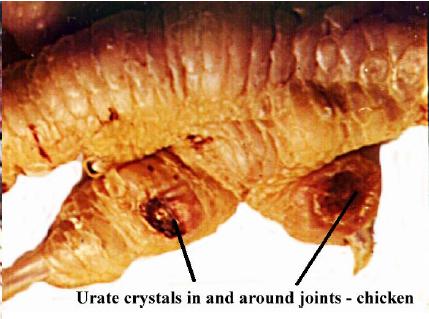
| 19:32, 21 August 2007 | Urate crystals joints.jpg (file) |  |
26 KB | Gross appearance of urate crystals in the joints. Pending permission from Brian Smyth. | 1 |
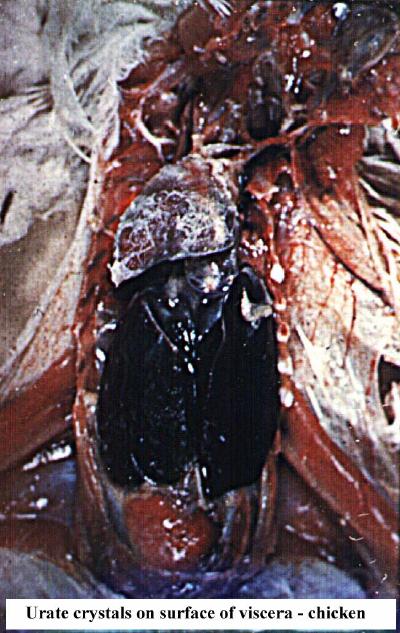
| 19:31, 21 August 2007 | Urate crystals viscera.jpg (file) |  |
59 KB | Gross appearance of urate crystals on the serous membranes. Pending permission from Brian Smyth. | 1 |
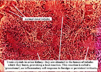
| 19:30, 21 August 2007 | Urate crystals kidney.jpg (file) |  |
50 KB | Histological appearance of urate crystals in the kidney. Pending permission from Brian Smyth. | 1 |
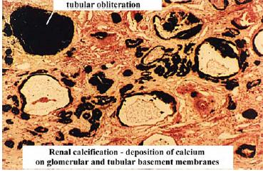
| 19:25, 21 August 2007 | Renal calcification.jpg (file) |  |
34 KB | Histological appearance of calcification in the kidney. Pending permission from Brian Smyth. | 1 |
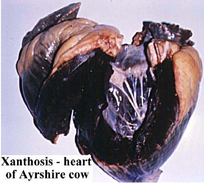
| 18:18, 21 August 2007 | Lipofuscin xanthosis.jpg (file) |  |
30 KB | Xanthosis in the heart of an Ayrshire cow. Pending permission from Brian Smyth. | 1 |
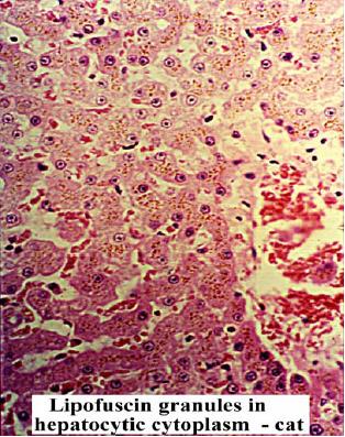
| 18:17, 21 August 2007 | Lipofuscin granules 2.jpg (file) |  |
45 KB | Lipofuscin granules in the cytoplasm of hepatocytes in the cat. Pending permission from Brian Smyth. | 1 |
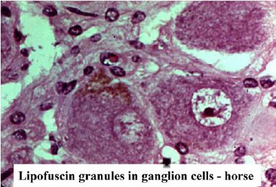
| 18:16, 21 August 2007 | Lipofuscin granules.jpg (file) |  |
28 KB | Histological appearance of lipofuscine granules in ganglion cells of the horse. Pending permission from Brian Smyth. | 1 |
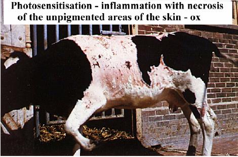
| 18:13, 21 August 2007 | Photosensitisation.jpg (file) |  |
37 KB | Inflammation of the skin due to photosensitisation in the ox. Pending permission from Brian Smyth. | 1 |
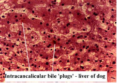
| 18:10, 21 August 2007 | Jaundice bile plugs.jpg (file) |  |
28 KB | Intracanalicular "bile plugs" in the liver of the dog due to jaundice. Pending permission from Brian Smyth. | 1 |
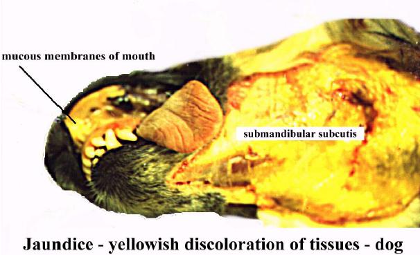
| 18:09, 21 August 2007 | Jaundice discolouration.jpg (file) |  |
38 KB | Yellow discolouration of the mucouse membranse and the mandibular subcutis due to jaundice in the dog. Pending permission from Brian Smyth. | 1 |
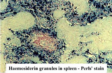
| 17:02, 21 August 2007 | Haemosiderin prussian.jpg (file) |  |
36 KB | Section stained with Prussian Blue to show haemosiderin. Pending permission from Brian Smyth. | 1 |
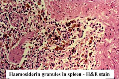
| 14:57, 21 August 2007 | Haemosiderin.jpg (file) |  |
35 KB | Histological appearance of haemosidering granules. Pending permission from Brian Smyth. | 1 |
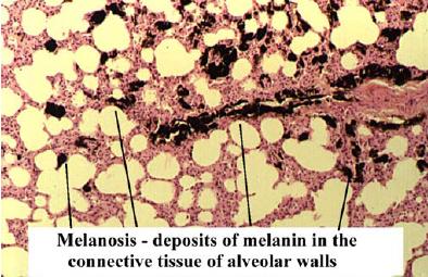
| 13:23, 21 August 2007 | Melanosis histo.jpg (file) |  |
34 KB | Histological appearance of melanosis. Pending permission from Brian Smyth. | 1 |
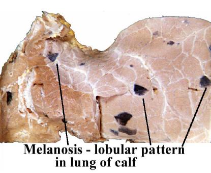
| 13:23, 21 August 2007 | Melanosis gross.jpg (file) |  |
27 KB | Gross appearance of melanosis. Pending permission from Brian Smyth. | 1 |
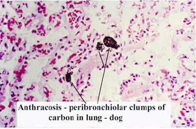
| 12:27, 21 August 2007 | Anthracosis histo.jpg (file) |  |
27 KB | Histological appearance of anthracosis. Pending permission from Brian Smyth. | 1 |
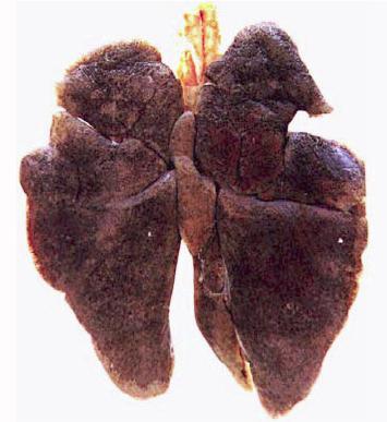
| 12:26, 21 August 2007 | Anthracosis gross.jpg (file) |  |
23 KB | Gross appearance of antrhacosis in the lungs of a dog. Pending permission from Brian Smyth. | 1 |
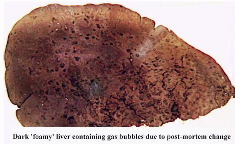
| 12:03, 21 August 2007 | Foamy liver.jpg (file) |  |
27 KB | Appearance of a foamy lover due to putrefaction. Pending permission from Brian Smyth. | 1 |
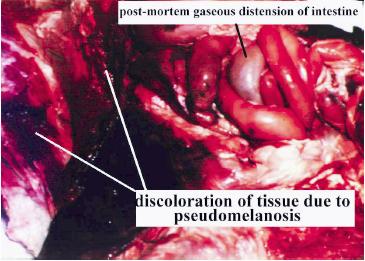
| 12:01, 21 August 2007 | Pseudomelanosis.jpg (file) |  |
24 KB | Appearance of pseudomelanosis. Pending permission from Brian Smyth. | 1 |
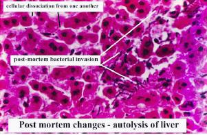
| 10:35, 21 August 2007 | Autolysis of liver.jpg (file) |  |
21 KB | Histological appearance of autolysis in the liver. Pending permission from Brian Smyth. | 1 |
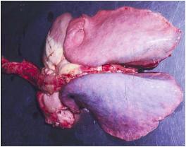
| 09:58, 21 August 2007 | Hypostatic congestion.jpg (file) |  |
11 KB | Hypostatic congestion in the lungs. Pending permission from Brian Smyth. | 1 |
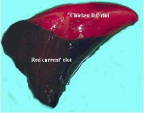
| 09:51, 21 August 2007 | Clot.jpg (file) |  |
8 KB | A post-mortem clot, showing the upper "chicken-fat" clot and the lower "redcurrant-jelly" clot. Pending permission from Brian Smyth. | 1 |
| 21:30, 20 August 2007 | Tuberculosis granuloma.jpg (file) |  |
29 KB | Tuberculosis granuloma. Pending permission from Brian Smyth. | 1 |
| 21:27, 20 August 2007 | Calcification.jpg (file) |  |
19 KB | Histological appearance of calcification. Pending permission from Brian Smyth. | 1 |
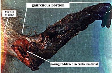
| 21:25, 20 August 2007 | Monkey gangrene 2.jpg (file) |  |
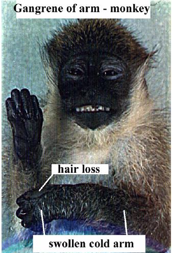
27 KB | Wet gangrene in a monkey (2). Pending permission from Brian Smyth. | 1 |
| 21:23, 20 August 2007 | Monkey gangrene 1.jpg (file) |  |
41 KB | Wet gngrene in a monkey (1). Pending permission form Brian Smyth. | 1 |
| 21:20, 20 August 2007 | Necrosis histo.jpg (file) |  |
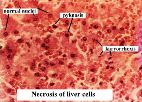
37 KB | Histological appearance of necrosis. Pending permission from Brian Smyth. | 1 |