File list
Jump to navigation
Jump to search
This special page shows all uploaded files.
| Date | Name | Thumbnail | Size | User | Description | Versions |
|---|---|---|---|---|---|---|
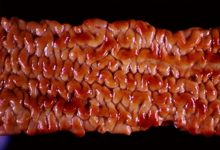
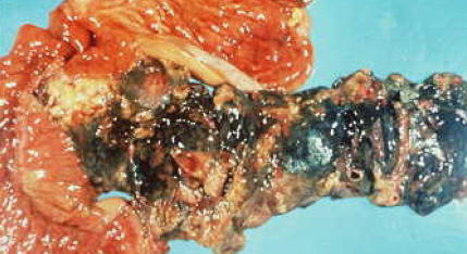
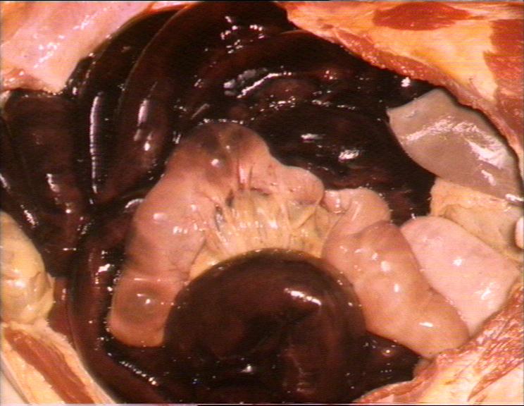
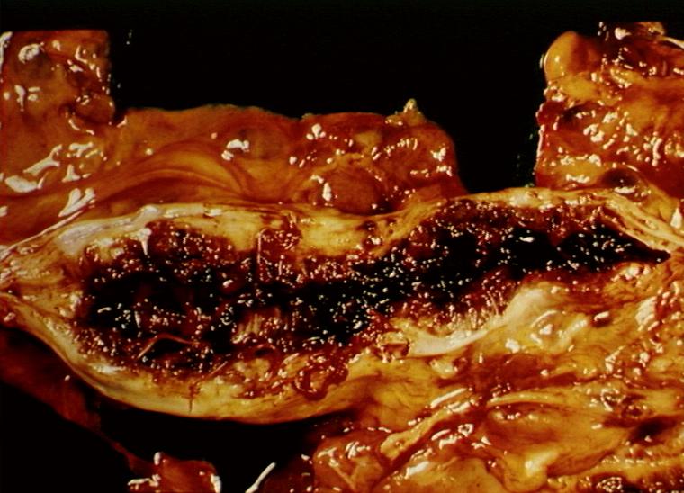
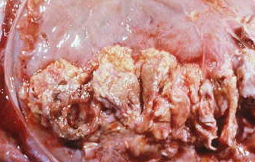
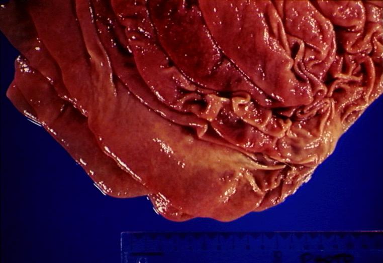
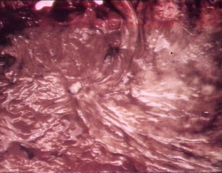
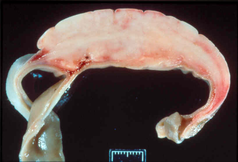
| 20:19, 14 August 2007 | Johnes disease proliferative ileitis.jpg (file) |  |
63 KB | Lizzies | Ileum opened: proliferative ileitis in Johne's disease. | 1 |
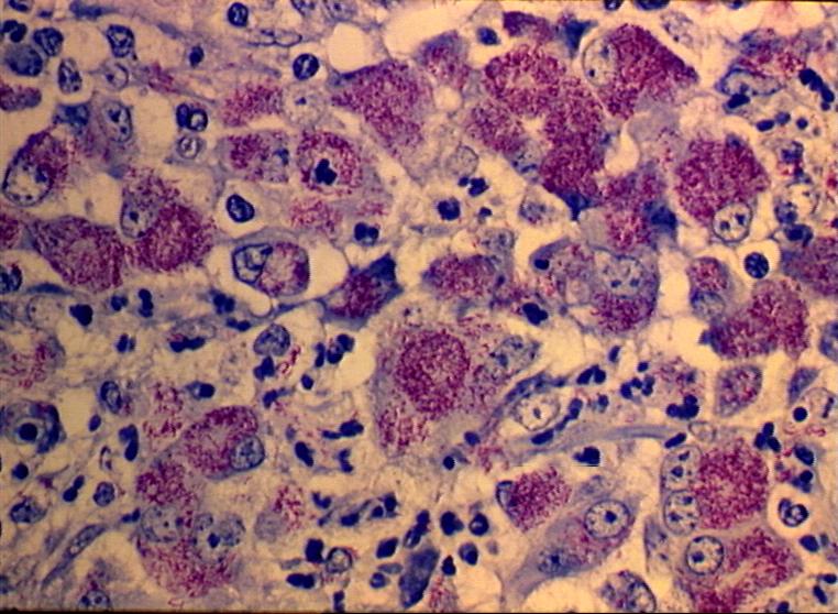
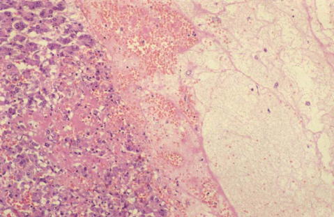
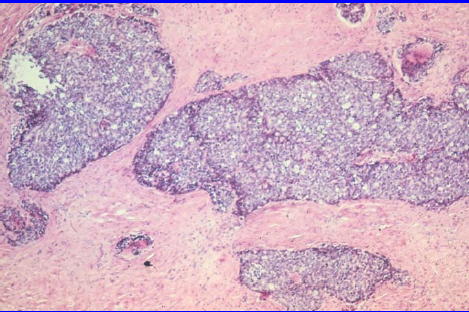
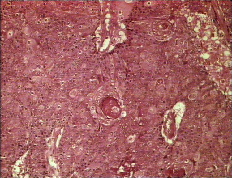
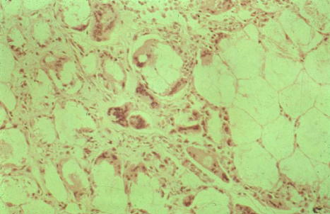
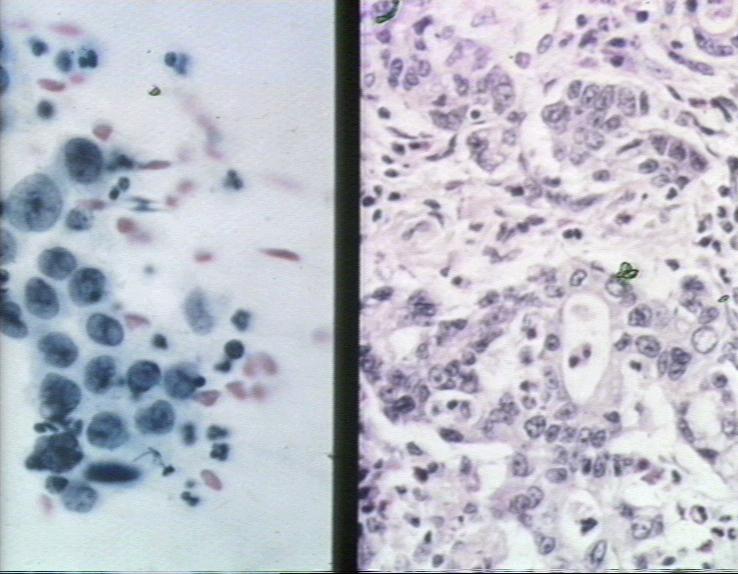
| 20:17, 14 August 2007 | Johnes disease histological.jpg (file) |  |
100 KB | Lizzies | Johne's disease in the ileum- histological. | 1 |
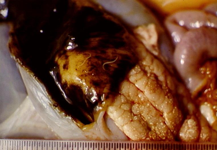
| 19:45, 14 August 2007 | Trichuris ovis.jpg (file) |  |
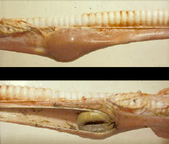
55 KB | Lizzies | Trichuris ovis in caecum. | 1 |
| 19:39, 14 August 2007 | Trichuris vulpis caecum comparative.jpg (file) |  |
69 KB | Lizzies | Dog caecum: left normal, right with mucosal hyperplasia and inflammation caused by Trichuris vulpis . | 1 |
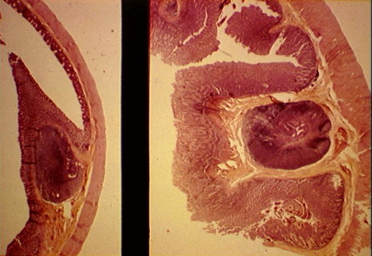
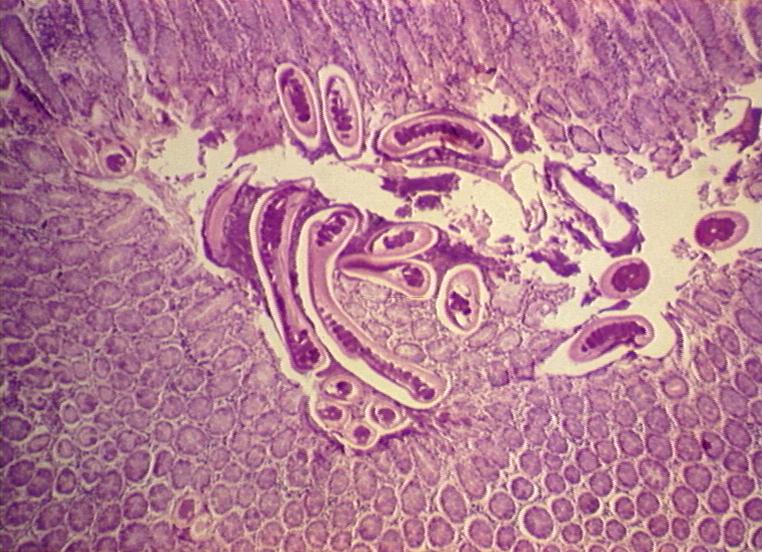
| 19:38, 14 August 2007 | Trichuris vulpis caecum.jpg (file) |  |
100 KB | Lizzies | Caecum showing hyperplastic mucosa and sections of Trichuris vulpis (dog whipworm) and eggs . | 1 |
| 11:24, 14 August 2007 | Bucket and spade.jpg (file) |  |
14 KB | Gillybean | Free of copyright image | 1 |
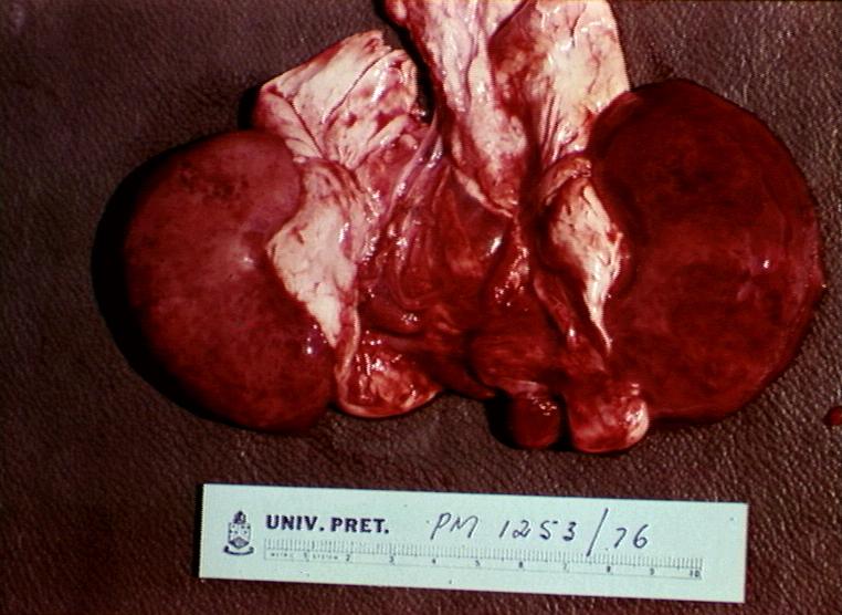
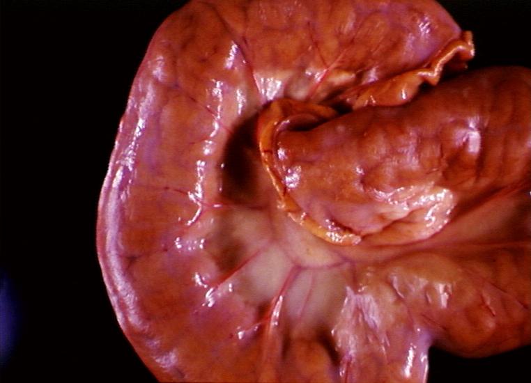
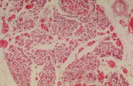
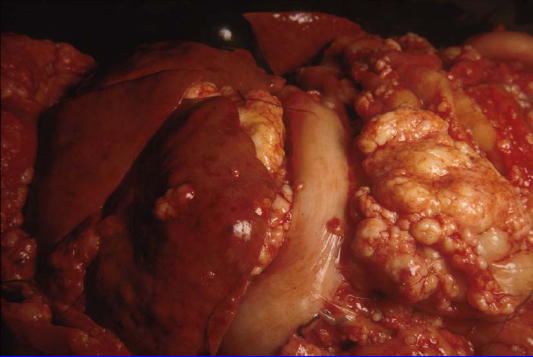
| 21:19, 13 August 2007 | Pulpy kidney gross.jpg (file) |  |
70 KB | Lizzies | Renal haemorrhage and nephrosis: pulpy kidney in enterotoxaemia. | 1 |
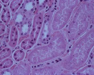
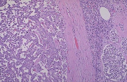
| 21:16, 13 August 2007 | Pulpy kidney disease.jpg (file) |  |
15 KB | Lizzies | Abrupt transition between viable and necrotic renal tubules in pulpy kidney disease. | 1 |
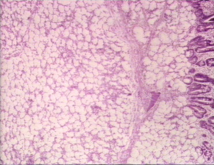
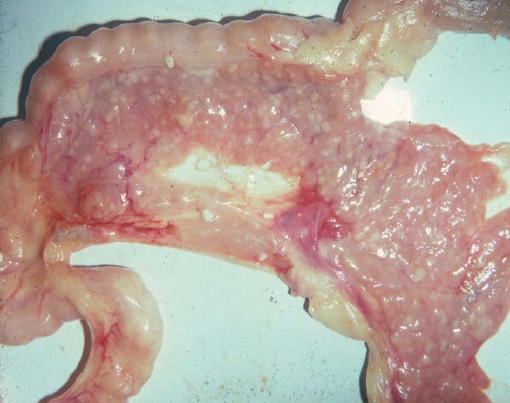
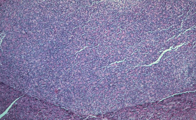
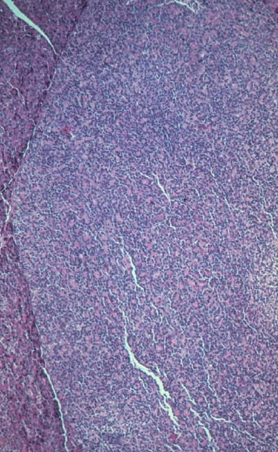
| 21:06, 13 August 2007 | Johnes disease proliferative enteritis.jpg (file) |  |
50 KB | Lizzies | Johnes disease - proliferative enteritis. | 1 |
| 10:55, 13 August 2007 | Gill2.jpg (file) |  |
14 KB | Gillybean | Me | 1 |
| 16:24, 10 August 2007 | Atresia ani PM.jpg (file) |  |
94 KB | Srhind | 1 | |
| 13:34, 10 August 2007 | Beta cell carcinoma.jpeg (file) |  |
48 KB | Bara | 1 | |
| 13:34, 10 August 2007 | Insulinoma.jpeg (file) |  |
56 KB | Bara | 1 | |
| 11:25, 10 August 2007 | Acute pancreatic necrosis.jpeg (file) |  |
30 KB | Bara | 1 | |
| 11:25, 10 August 2007 | Acute haemorrhagic pancreatitis.jpeg (file) |  |
37 KB | Bara | 1 | |
| 11:24, 10 August 2007 | Brunner gland adenoma.jpg (file) |  |
75 KB | Lizzies | Adenoma of brunners gland (duodenum). | 1 |
| 11:22, 10 August 2007 | Pancreatic hypoplasia micro.jpeg (file) |  |
34 KB | Bara | 1 | |
| 11:21, 10 August 2007 | Pancreatic hypoplasia.jpeg (file) |  |
10 KB | Bara | 1 | |
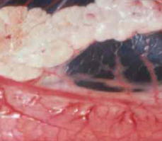
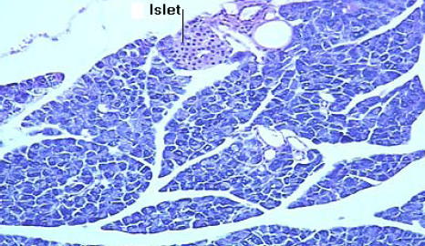
| 11:20, 10 August 2007 | Islet.jpeg (file) |  |
48 KB | Bara | 1 | |
| 11:19, 10 August 2007 | Pancreatic nodular hyperplasia.jpeg (file) |  |
36 KB | Bara | 1 | |
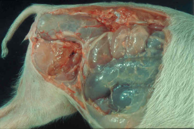
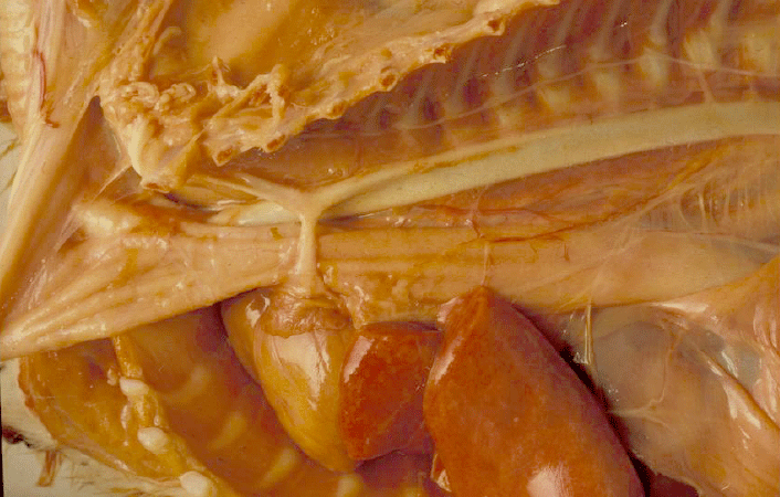
| 11:15, 10 August 2007 | Infaction of the small bowel.jpg (file) |  |
65 KB | Lizzies | Infaction of the small bowel. | 1 |
| 11:09, 10 August 2007 | Carcinoma micro.jpeg (file) |  |
44 KB | Bara | 1 | |
| 11:09, 10 August 2007 | Carcinoma gross.jpeg (file) |  |
30 KB | Bara | 1 | |
| 11:06, 10 August 2007 | Carcinoma.jpeg (file) |  |
30 KB | Bara | 1 | |
| 11:05, 10 August 2007 | Adenoma2.jpeg (file) |  |
82 KB | Bara | 1 | |
| 11:00, 10 August 2007 | Strongylus vulgaris.jpg (file) |  |
78 KB | Lizzies | Thrombosis of cranial mesenteric artery caused by Strongylus vulgaris larvae. | 1 |
| 10:52, 10 August 2007 | Perianal gland adenoma.jpg (file) |  |
51 KB | Lizzies | Gross appearance of a perianal gland adenoma. | 1 |
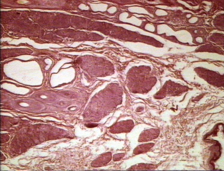
| 10:51, 10 August 2007 | Perianal gland adenoma histopath.jpg (file) |  |
100 KB | Lizzies | Histological appearance of a perianal gland adenoma. | 1 |
| 10:51, 10 August 2007 | Adenoma.jpeg (file) |  |
83 KB | Bara | 1 | |
| 10:51, 10 August 2007 | Normal perianal gland.jpg (file) |  |
97 KB | Lizzies | Histological appearance of a normal perianal gland. | 1 |
| 10:49, 10 August 2007 | Chronic pancreatitis.jpeg (file) |  |
41 KB | Bara | 1 | |
| 10:43, 10 August 2007 | Acute interstitial pancreatitis.jpeg (file) |  |
30 KB | Bara | 1 | |
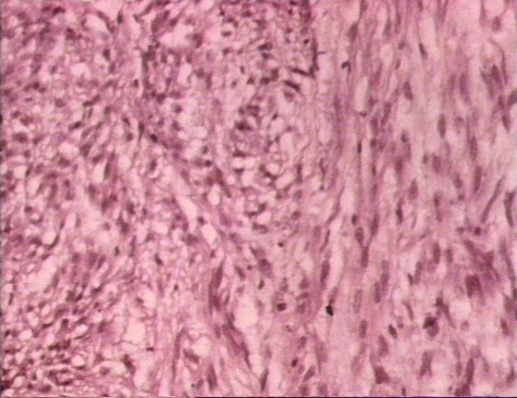
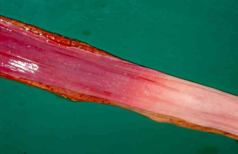
| 19:16, 9 August 2007 | Leiomyoma.jpg (file) |  |
67 KB | Lizzies | Histological appearance of leiomyoma. | 1 |
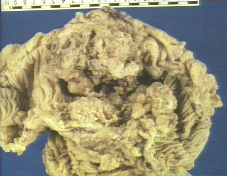
| 19:07, 9 August 2007 | Ostertagiasis.jpg (file) |  |
57 KB | Lizzies | Ostertagiosis caused by Ostertagia ostertagi: abomasum showing oedema and inflamation of fundic folds. | 1 |
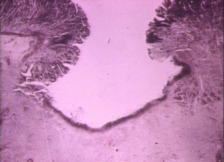
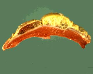
| 19:04, 9 August 2007 | Gastric ulcer histopath.jpg (file) |  |
55 KB | Lizzies | Histological section through a gastric ulcer. | 1 |
| 19:03, 9 August 2007 | Gastric ulcer.jpg (file) |  |
65 KB | Lizzies | Gross appearance of a gastric ulcer. | 1 |
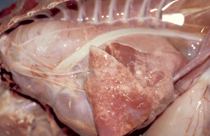
| 18:52, 9 August 2007 | Traumatic pericarditis.jpg (file) |  |
7 KB | Lizzies | 1 | |
| 18:46, 9 August 2007 | Oesophageal bloat line.jpg (file) |  |
58 KB | Lizzies | 1 | |
| 18:39, 9 August 2007 | Adenocarcinoma stomach.jpg (file) |  |
69 KB | Lizzies | 1 | |
| 18:39, 9 August 2007 | Adenocarcinoma stomach histopath2.jpg (file) |  |
64 KB | Lizzies | 1 | |
| 18:24, 9 August 2007 | Abomasal lymphoma.jpg (file) |  |
68 KB | Lizzies | 1 | |
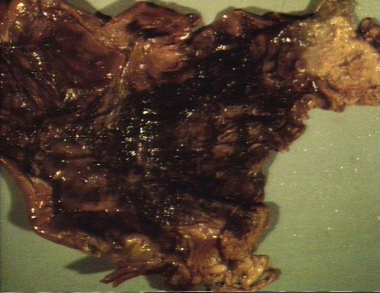
| 16:21, 9 August 2007 | Haemorrhagic gastritis.jpg (file) |  |
63 KB | Lizzies | 1 | |
| 13:08, 9 August 2007 | Mindmap.pdf (file) | 23 KB | Brian | PDF Mind Map | 1 | |
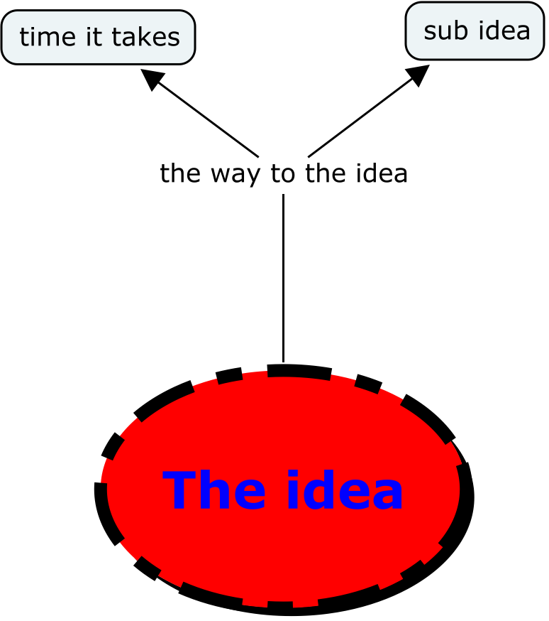
| 10:45, 9 August 2007 | Mindmap.png (file) |  |
41 KB | Brian | this is my idea | 1 |
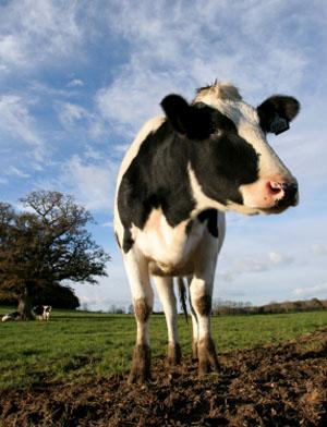
| 16:28, 7 August 2007 | Cow2.jpg (file) |  |
24 KB | Lizzies | 1 | |
| 10:41, 7 August 2007 | Bovimpaction.gif (file) |  |
60 KB | Alexm1983 | Impaction in the bovine oesophagus - Picture courtesy of Alun Williams | 1 |
| 10:36, 7 August 2007 | Megaoes.gif (file) |  |
215 KB | Alexm1983 | Megaoesophagus - Picture courtesy of Alun Williams | 1 |
| 10:32, 7 August 2007 | Praa.gif (file) |  |
237 KB | Alexm1983 | Dextra-aorta - Picture courtesy of Alun Williams (RVC) | 1 |
| 00:35, 7 August 2007 | CmapTools - Home Page Cmap.pdf (file) | 280 KB | Alexm1983 | 1 | ||
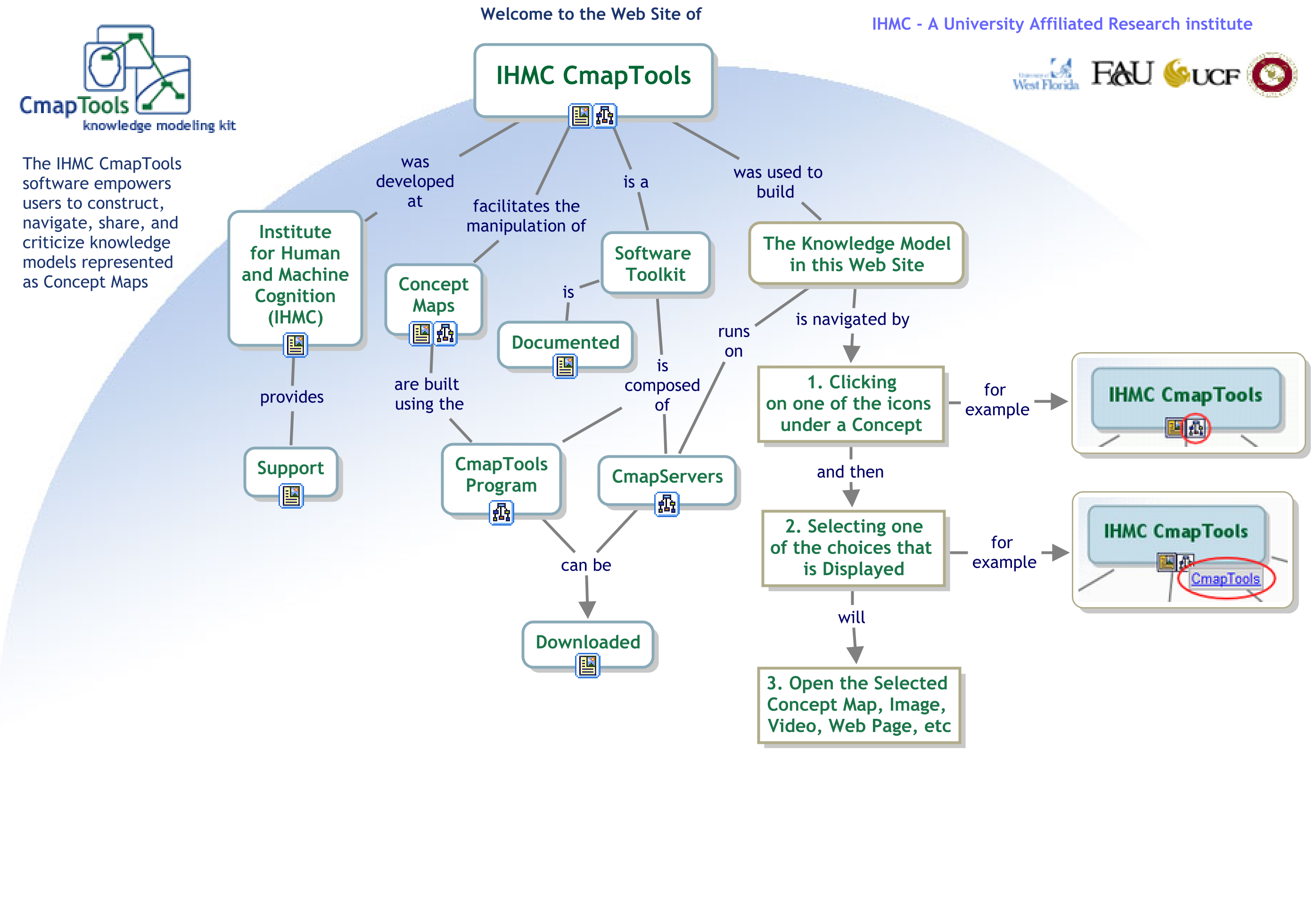
| 00:34, 7 August 2007 | CmapTools - Home Page Cmap.png (file) |  |
390 KB | Alexm1983 | 1 |