Unused files
The following files exist but are not embedded in any page. Please note that other web sites may link to a file with a direct URL, and so may still be listed here despite being in active use.
Showing below up to 50 results in range #401 to #450.
View (previous 50 | next 50) (20 | 50 | 100 | 250 | 500)
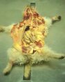
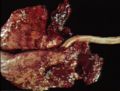
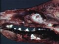
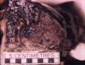
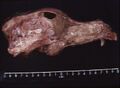 Lymphoma in nasal cavity.jpg 748 × 550; 50 KB
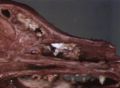
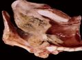
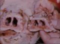
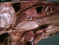 Lymphoma in pharynx.jpg 750 × 566; 71 KB
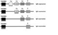 Immunoglobulin heavy chain genes.jpg 647 × 386; 29 KB
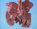
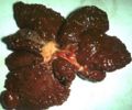 01 - Nodular liver failure.JPG 394 × 328; 24 KB
 01 - Jaundice liver failure.JPG 408 × 518; 29 KB
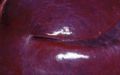
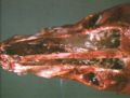
 03 - Photosensitisation.JPG 406 × 265; 31 KB
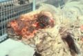 04 - Photosensitisation sheep.JPG 371 × 249; 19 KB
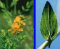 05 - st johns wort.JPG 362 × 297; 20 KB
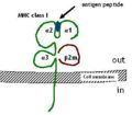 MHC I.jpg 381 × 333; 19 KB
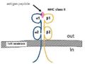 MHC II.jpg 419 × 315; 19 KB
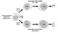 TH development.jpg 651 × 381; 32 KB
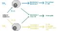 TH function.jpg 734 × 399; 33 KB
 06 - portosystemic shunting.JPG 403 × 248; 30 KB
 07 - portosystemic shunting.JPG 529 × 483; 43 KB
 08 - acute liver damage.JPG 433 × 281; 20 KB
 09 - necrosis of the liver.JPG 411 × 272; 22 KB
 10 - random focal.JPG 555 × 349; 23 KB
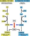 Complement activation.jpg 576 × 701; 56 KB
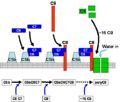 Membrane attack complex formation.jpg 576 × 491; 41 KB
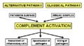 Complement activity.jpg 509 × 282; 29 KB
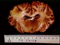 Focal leukoencephalomalacia.jpg 768 × 576; 55 KB
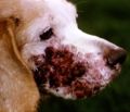 Cutaneous lymphosarcoma.jpg 664 × 572; 57 KB
 Miliary tuberculosis.jpg 768 × 576; 51 KB
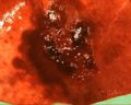 Consolidation and haemorrhage lung.jpg 320 × 256; 16 KB
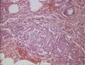
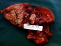
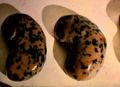 Kidney melanosis.jpg 760 × 552; 57 KB
 Comparative kidneys; cambridge.jpg 644 × 197; 25 KB
 Cambridge comparative.jpg 1,502 × 1,127; 9 KB
 Picture1.png 1,502 × 1,127; 2 KB
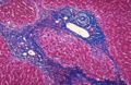
 11 - zonal necrosis.JPG 512 × 323; 60 KB
 12 - ICH.JPG 376 × 225; 22 KB
 13 - massive necrosis.JPG 478 × 355; 40 KB
 14 - fibrosis.JPG 400 × 261; 46 KB
 Metastatic fibrosarcoma.jpg 738 × 574; 64 KB
 Metastatic sweat gland carcinoma.jpg 320 × 245; 19 KB
 Mucoid rhinitis.jpg 746 × 568; 67 KB
 Multiple pulmonary abscesses.jpg 752 × 572; 65 KB
 Aspergillosis in nasal cavity.jpg 744 × 546; 55 KB
 Necrotising laryngitis.jpg 752 × 550; 53 KB
 Nasal cavity carcinoma.jpg 752 × 564; 68 KB
 Nasopharyngeal fistula.jpg 750 × 566; 57 KB
 Oedema and chondritis in larynx of sheep.jpg 748 × 572; 73 KB
 Pyogranulomatous lungs due to Rhodococcus Equi.jpg 750 × 566; 58 KB
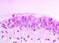 Trachea epithelium.jpg 756 × 562; 67 KB
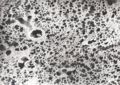 Lung scanning electron micrograph.jpg 1,138 × 809; 228 KB
 Bronchus.jpg 744 × 561; 46 KB
 Bronchus, bronchiole, blood vessel.jpg 1,434 × 754; 177 KB
 Alveoli.jpg 1,050 × 690; 105 KB
 Alveolar macrophages.jpg 754 × 563; 47 KB
 Segmental pulmonary infarction.jpg 746 × 570; 74 KB
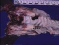
 Severe atrophic rhinitis.jpg 748 × 550; 55 KB


