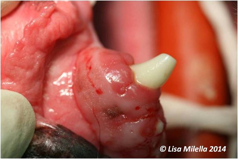
File:Gingivoplasty step 1.jpg
Revision as of 15:13, 10 April 2014 by Bara (talk | contribs) ({{Information |Description ={{en|1=Gingivoplasty step 1 - pocket depths are measured with a graduated periodontal probe. The probe is withdrawn from the pocket and held against the outer surface of the gingiva to show the depth of the pocket and a blee)

Size of this preview: 800 × 535 pixels. Other resolutions: 320 × 214 pixels | 1,186 × 793 pixels.
Original file (1,186 × 793 pixels, file size: 66 KB, MIME type: image/jpeg)
Summary
| Description |
English: Gingivoplasty step 1 - pocket depths are measured with a graduated periodontal probe. The probe is withdrawn from the pocket and held against the outer surface of the gingiva to show the depth of the pocket and a bleeding point made. This is repeated along the circumference of the tooth.
|
|---|---|
| Date |
2014 |
| Source |
Lisa Milella |
| Author |
Lisa Milella |
| Permission (Reusing this file) |
See below |
Licensing

|
The author of this file has reserved all rights. Permission needs to be obtained from the author before its use. |
File history
Click on a date/time to view the file as it appeared at that time.
| Date/Time | Thumbnail | Dimensions | User | Comment | |
|---|---|---|---|---|---|
| current | 15:13, 10 April 2014 |  | 1,186 × 793 (66 KB) | Bara (talk | contribs) | {{Information |Description ={{en|1=Gingivoplasty step 1 - pocket depths are measured with a graduated periodontal probe. The probe is withdrawn from the pocket and held against the outer surface of the gingiva to show the depth of the pocket and a blee |
You cannot overwrite this file.
File usage
The following page uses this file: