Unused files
Jump to navigation
Jump to search
The following files exist but are not embedded in any page. Please note that other web sites may link to a file with a direct URL, and so may still be listed here despite being in active use.
Showing below up to 50 results in range #811 to #860.
View (previous 50 | next 50) (20 | 50 | 100 | 250 | 500)
- Dissacharidase.jpg 1,416 × 745; 136 KB
- LH Hassalls Corpuscle Histology.jpg 825 × 647; 177 KB
- LH Bursa Cloaca Photo.jpg 601 × 272; 52 KB
- LH Bursa High Histology.jpg 533 × 685; 195 KB
- LH Bursa Low Histology.jpg 829 × 645; 133 KB
- LH Bursa Photo.jpg 668 × 441; 61 KB
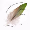
- Parts of the Feather.jpg 599 × 600; 36 KB
- LH Ileal Peyer's Patch Diagram.jpg 1,416 × 724; 124 KB
- LH Ileal Peyer's Patch Histology.jpg 821 × 432; 125 KB
- LH Ileal Peyer's Patch Low Histology.jpg 796 × 484; 143 KB
- Gizzard Histology.jpg 640 × 500; 50 KB
- Shedding of Temporary Tooth.jpg 720 × 477; 63 KB
- Dog Dentition.jpg 640 × 400; 32 KB
- Tooth Radiograph.jpg 640 × 400; 15 KB
- Feline Dentition.jpg 640 × 400; 28 KB
- Rabbit Teeth.jpg 640 × 400; 23 KB
- Avian GIT.jpg 640 × 400; 36 KB
- Bursa of Fabricus.jpg 640 × 400; 23 KB
- Avian Cloaca Diagram.jpg 845 × 634; 71 KB
- WIKIVETspinalcord1.jpg 680 × 431; 56 KB
- WIKIVETspinalcord2.jpg 700 × 410; 49 KB
- WIKIVETspinalcord3.jpg 674 × 360; 32 KB
- WIKIVETcerebrum.jpg 631 × 377; 44 KB
- WIKIVETcerebellum.jpg 650 × 378; 35 KB
- Carotidretesheep.jpg 1,130 × 551; 52 KB
- WIKIVETmyelinatednervesinsmallfibreinlongittudinlsection.jpg 683 × 346; 32 KB
- WIKIVETmyleinatednerveinsmallfibre.jpg 677 × 360; 34 KB
- WIKIVETmyeinatednerveinsmallfibrelipdstained.jpg 676 × 347; 37 KB
- WIKIVETautonomicganglion.jpg 663 × 394; 49 KB
- WIKIVETdorsalrootganglion.jpg 652 × 401; 53 KB
- Smalllargeintestine.jpg 448 × 678; 53 KB
- Small&largeintestine.jpg 448 × 678; 53 KB
- Opendogcaecum.jpg 448 × 676; 51 KB
- Illustration dog descending colon.jpg 447 × 678; 38 KB
- Stomach Anatomy 1.jpg 640 × 622; 59 KB
- Stomach Anatomy 2.jpg 640 × 544; 49 KB
- Stomach Anatomy 3.jpg 640 × 528; 50 KB
- Margo Plicatus.jpg 663 × 494; 37 KB
- Formation of Bile Acids.jpg 844 × 633; 57 KB
- Pig Liver Topography.jpg 640 × 414; 33 KB
- Canine Liver Topography.jpg 733 × 400; 27 KB
- Recto-Anal Junction.jpg 764 × 568; 84 KB
- Cockatiel.jpg 984 × 733; 61 KB
- Vitreous-Retina.jpg 640 × 400; 23 KB
- Detailed globe.jpg 640 × 400; 42 KB
- Pancreas Sheep.jpg 1,448 × 973; 195 KB
- WIKIVETformationofneuraltissue.jpg 793 × 447; 31 KB
- WIKIVETneuraltubeformation.jpg 562 × 273; 16 KB
- Sheep Pancreas.jpg 704 × 569; 70 KB
- LH Spleen Rat Histology.jpg 722 × 362; 74 KB


