Search results
Jump to navigation
Jump to search
Page title matches

File:Vibrio parahaemolyticus- An emerging foodborne pathogen-A Review.pdf (222 KB) - 10:58, 24 December 2011
Page text matches
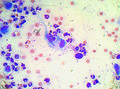
File:Cytology 17a.JPG ...Publishing]], as part of the [[OVAL]]. This is an smear of an aspirate of an interdigital mass showing moderate to marked pyogranulomatous inflammation(828 × 616 (740 KB)) - 18:07, 28 September 2011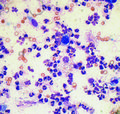
File:Cytology 17b.JPG ...Publishing]], as part of the [[OVAL]]. This is an smear of an aspirate of an interdigital mass showing moderate to marked pyogranulomatous inflammation(717 × 680 (874 KB)) - 18:09, 28 September 2011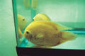
File:Ornamental Fish 11.jpg ...[Manson|Manson Publishing]], as part of the [[OVAL]]. This is an image of an adult gold severum infected with ''Ichthyophthirius multifiliis''.(500 × 330 (225 KB)) - 18:51, 22 September 2011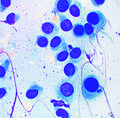
File:Cytology 09.JPG ...[Manson|Manson Publishing]], as part of the [[OVAL]]. This is an smear of an FNA from a histiocytoma.(789 × 778 (774 KB)) - 15:57, 27 September 2011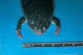
File:Rep 08.jpg ...[Manson|Manson Publishing]], as part of the [[OVAL]]. This is an image of an African clawed frog with aeromoniasis(476 × 313 (98 KB)) - 20:41, 15 February 2012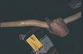
File:Rep 22.jpg ...[Manson|Manson Publishing]], as part of the [[OVAL]]. This is an image of an Australian taipan snake with cardiomyopathy.(460 × 300 (48 KB)) - 20:51, 15 February 2012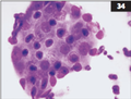
File:Cytology 07.png ...[Manson|Manson Publishing]], as part of the [[OVAL]]. This is an smear of an aspirate from a perianal adenoma.(404 × 308 (120 KB)) - 14:54, 29 September 2011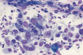
File:Cytology 04.jpg ...[Manson|Manson Publishing]], as part of the [[OVAL]]. This is an smear of an FNA from a Golden Retriever with sarcoma.(500 × 328 (192 KB)) - 07:39, 29 August 2011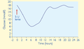
File:Feline Medicine 03.png ...[[OVAL]]. This is an image of a graph showing blood glucose results from an insulin resistant diabetic cat.(587 × 344 (19 KB)) - 07:45, 2 October 2011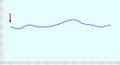
File:Feline Medicine 03.jpg ...[[OVAL]]. This is an image of a graph showing blood glucose results from an insulin resistant diabetic cat.(277 × 151 (559 KB)) - 13:24, 29 September 2011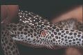
File:Rep 21.jpg ...[Manson|Manson Publishing]], as part of the [[OVAL]]. This is an image of an a collared lizard with ''Herstiella trombidiiformis'' infestation.(438 × 291 (75 KB)) - 20:50, 15 February 2012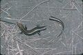
File:Rep 24.jpg ...[Manson|Manson Publishing]], as part of the [[OVAL]]. This is an image of an insectivorous skink which has shed its tail(456 × 305 (112 KB)) - 20:52, 15 February 2012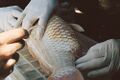
File:Ornamental Fish 02a.jpg ...the [[OVAL]]. This is an image of a mature female tancho kohaku koi with an ovarian prolapse through the genital pore.(500 × 334 (197 KB)) - 19:30, 19 September 2011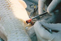
File:Ornamental Fish 02b.jpg ...the [[OVAL]]. This is an image of a mature female tancho kohaku koi with an ovarian prolapse through the genital pore.(500 × 333 (189 KB)) - 19:32, 19 September 2011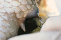
File:Ornamental Fish 02c.jpg ...the [[OVAL]]. This is an image of a mature female tancho kohaku koi with an ovarian prolapse through the genital pore.(500 × 334 (176 KB)) - 19:32, 19 September 2011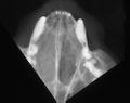
File:Feline Medicine 19.jpg ...[Manson|Manson Publishing]], as part of the [[OVAL]]. This is an image of an intra-oral nasal radiograph of a cat with nasal neoplasia.(500 × 397 (99 KB)) - 11:41, 25 August 2011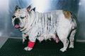
File:SA ST Sx 12.jpg ...[Manson|Manson Publishing]], as part of the [[OVAL]]. This is an image of an English bulldog with dystocia.(950 × 634 (318 KB)) - 14:28, 29 September 2011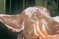
File:Soft Tissue Sx 17.jpg ...lishing]], as part of the [[OVAL]]. This is an image of a Weimaraner with an aural hematoma.(500 × 333 (173 KB)) - 13:50, 30 August 2011
File:Dogandwikivet.jpg ...the [[:File:Where's WikiVet.png|'Where's WikiVet' logo]] next to a dog, as an example for the 'Where's WikiVet' [[Photo Competition]]}}(448 × 745 (57 KB)) - 16:14, 30 March 2012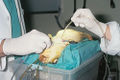
File:Ornamental Fish 16a.jpg ...e of a section from the gill of a koi being prepared for surgery to remove an abdominal mass.(500 × 334 (209 KB)) - 16:50, 20 September 2011


