Uploads by Svycrnj
This special page shows all uploaded files.
| Date | Name | Thumbnail | Size | Description | Versions |
|---|---|---|---|---|---|
| 14:57, 5 September 2010 | Cooperia oncophora L3.jpg (file) |  |
30 KB | {{Information |Description=Image fo L3 C. oncophora |Source=RVC Veterinary Parasitology online resource |Date= |Author= |Permission=See below |Other_versions= }} | 1 |
| 17:33, 8 August 2010 | Ascaris suum egg.jpg (file) | 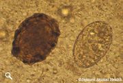 |
20 KB | {{Information |Description=''A. suum'' egg |Source=RVC/FAO Guide to veterinary parasitology. http://www.rvc.ac.uk/Review/Parasitology/pigEggs/ascaris.htm |Date= |Author= Janssen Animal Health |Permission= RVC permission granted |other_versions= }} | 1 |
| 17:12, 8 August 2010 | Female nematode xsection.jpg (file) | 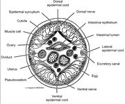 |
82 KB | {{Information |Description=Cross section of a female nematode |Source=http://biodidac.bio.uottawa.ca/thumbnails/filedet.htm?File_name=nema006b&File_type=cdr |Date= |Author=I. Livingstone |Permission=See Below |other_versions= }} | 1 |
| 16:21, 8 August 2010 | Nematode muscle.gif (file) | 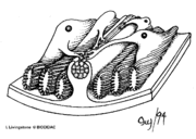 |
29 KB | {{Information |Description=Diagram of nematode muscle arrangement and innervation |Source=http://biodidac.bio.uottawa.ca/thumbnails/filedet.htm?File_name=NEMA005B&File_type=GIF |Date= |Author=I. Livingstone |Permission=See Below |other_versions= }} | 1 |
| 15:55, 8 August 2010 | Nematode pharynx.jpg (file) | 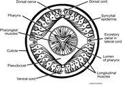 |
81 KB | {{Information |Description=Cross section of a nematode at the level of the pharynx |Source=http://biodidac.bio.uottawa.ca/thumbnails/catquery.htm?Kingdom=Animalia&phylum=Nematoda |Date= |Author= |Permission=See Below |other_versions= }} | 1 |
| 15:46, 8 August 2010 | Nematode copulation.jpg (file) | 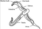 |
42 KB | {{Information |Description=Diagram of nematode copulation |Source=http://biodidac.bio.uottawa.ca/thumbnails/catquery.htm?Kingdom=Animalia&phylum=Nematoda |Date= |Author= |Permission= |other_versions= }} | 1 |
| 13:56, 4 August 2010 | Amidostomum anseris egg.jpg (file) | 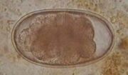 |
7 KB | {{Information |Description=An example of an ''A. anseris'' egg. |Source= http://heima.olivant.fo/~faroevet/fegvernisormur.htm |Date= |Author= Faroese Veterinary Service, Vardagota 85, FO-100 Torshavn, Faroe Islands. |Permission= Permission granted to W | 1 |
| 13:49, 4 August 2010 | Amidostomum anseris.jpg (file) |  |
27 KB | {{Information |Description=An example of an adult ''A. anseris'' worm |Source= http://heima.olivant.fo/~faroevet/fegvernisormur.htm |Date= |Author= Faroese Veterinary Service, Vardagota 85, FO-100 Torshavn, Faroe Islands. |Permission= Permission granted t | 1 |
| 11:34, 2 August 2010 | Toxocara canis adult.jpg (file) | 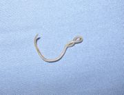 |
199 KB | {{Information |Description=''T. canis'' adult worm from a puppy |Source= Wikimedia commons |Date= 2006 |Author= Joel Mills |Permission= See Below |other_versions= }} | 1 |
| 16:55, 17 July 2010 | Rhipicephalus sanguineus.jpg (file) | 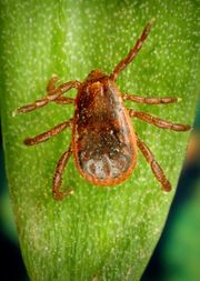 |
96 KB | {{Information |Description= Rhipicephalus sanguineus tick, also known as the brown dog tick |Source= CDC http://phil.cdc.gov |Date= 2005 |Author= James Gathany |Permission=Public domain }} | 1 |
| 12:07, 17 July 2010 | Ixodes holocyclus.jpg (file) | 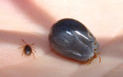 |
56 KB | {{Information |Description=''Ixodes holocylus'' ticks before and after a number of days feeding. |Source= Wikimedia commons |Date= 14 January 2009 |Author= Bjørn Christian Tørrissen |Permission=See below }} | 1 |
| 22:34, 16 July 2010 | Amblyomma americanum.jpg (file) | 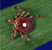 |
65 KB | {{Information |Description= The tick Amblyomma americanum (Lone Star tick) |Source= Wikimedia Commons |Date= n/a |Author= US Centers for Disease Control - Division of Vector Borne Infectious Diseases |Permission= Public Domain, US Federal Government work. | 1 |
| 12:06, 12 July 2010 | Trichuris Egg.jpg (file) | 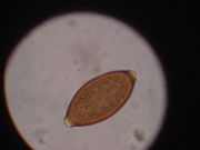 |
36 KB | Source: Wikimedia commons (http://commons.wikimedia.org/wiki/File:Whipworm_egg.JPG) Author: Joel Mills, 3/4/2006 | 1 |
| 12:54, 8 July 2010 | Dermacentor reticulatus.jpg (file) | 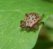 |
59 KB | Author: Rainer Altenkamp, Berlin Date: 25 August 2007 Lisence: Creative Commons Attribution-Share Alike 3.0 Unported Source: Wikimedia commons, taken on 8/7/2010 | 1 |
| 10:58, 8 July 2010 | Ixodidae life cycle.jpg (file) |  |
27 KB | Author: Centers for Disease Control and Prevention Public domain image. Wikimedia commons, taken on 8/7/2010 | 1 |
| 09:46, 8 July 2010 | Hpylori.jpg (file) | 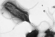 |
65 KB | H.pylori - © Yutaka Tsutsumi, M.D. Professor Department of Pathology Fujita Health University School of Medicine, Wikimedia Commons. Free use copyright, taken on 8/7/2010. | 1 |
| 10:57, 7 July 2010 | Mouthparts of Ixodes Holocyclus.jpg (file) | 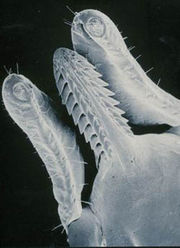 |
32 KB | Image used under the Creative Commons Attribution 3.0 Unported license. Author: Kevin Broady http://commons.wikimedia.org/wiki/File:Ixodholcapitulum.jpg, 7/7/2010 | 1 |
| 10:38, 7 July 2010 | Ixodes ricinus.jpg (file) | 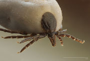 |
70 KB | Shared under the Creative Commons Attribution-Share Alike 2.5 Generic license. Author: Richard Bartz, 24 April 2009 http://commons.wikimedia.org/wiki/File:Ixodus_ricinus_5x.jpg, 7/7/2010 | 1 |
| 11:53, 5 July 2010 | Nickjackson.jpg (file) |  |
97 KB | Copyright Nicholas Jackson 2010 | 1 |