File list
This special page shows all uploaded files.
| Date | Name | Thumbnail | Size | User | Description | Versions |
|---|---|---|---|---|---|---|
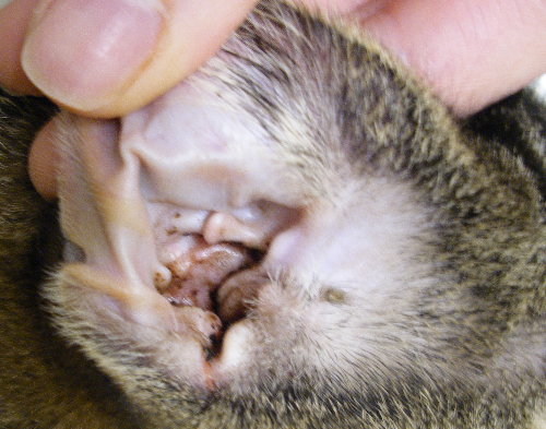
| 21:08, 20 February 2008 | Clinical Case 8 01.jpg (file) |  |
58 KB | Christina | 1 | |
| 21:17, 20 February 2008 | Clinical Case 8 02.jpg (file) |  |
26 KB | Christina | 1 | |
| 21:18, 20 February 2008 | Clinical Case 8 03.jpg (file) |  |
30 KB | Christina | 1 | |
| 21:07, 21 February 2008 | Clinical Case 9 01.jpg (file) |  |
32 KB | Christina | 1 | |
| 21:16, 21 February 2008 | Clinical Case 9 01b.jpg (file) |  |
32 KB | Christina | 1 | |
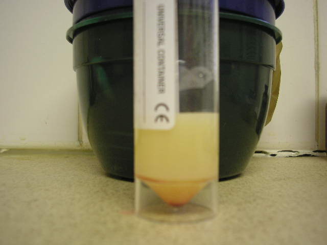
| 11:12, 25 February 2008 | Chyle.jpg (file) |  |
36 KB | Bara | 1 | |
| 20:47, 25 February 2008 | Clinical Case 10 01.jpg (file) |  |
55 KB | Christina | 1 | |
| 21:15, 25 February 2008 | Clinical Case 11 01.jpg (file) |  |
29 KB | Christina | 1 | |
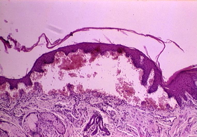
| 14:04, 28 February 2008 | Epidermolysis bullosa.jpg (file) |  |
224 KB | Bara | 1 | |
| 14:08, 28 February 2008 | Collagen dysplasia.jpg (file) |  |
165 KB | Bara | 1 | |
| 15:31, 3 March 2008 | Contagious ecthyma.jpg (file) |  |
48 KB | Bara | 1 | |
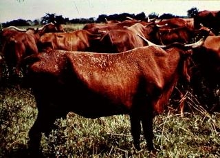
| 15:33, 3 March 2008 | Dermatophilosis in cow.jpg (file) |  |
67 KB | Bara | 1 | |
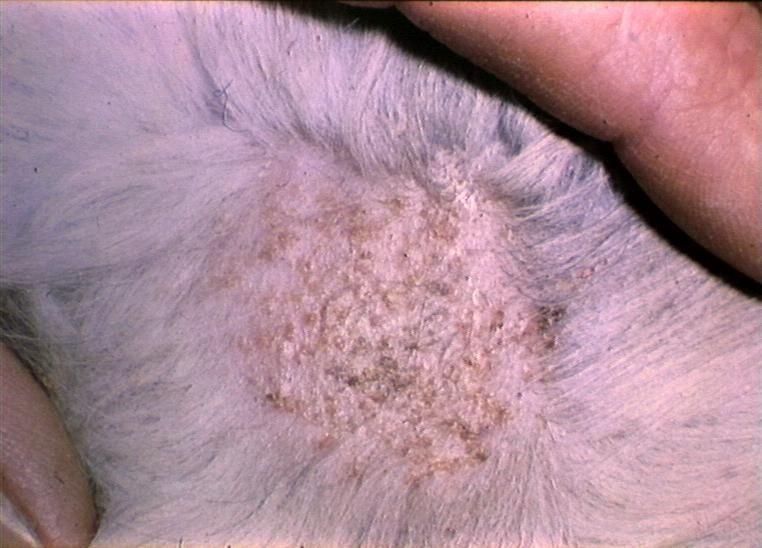
| 15:36, 3 March 2008 | Microsporum canis lesions.jpg (file) |  |
187 KB | Bara | 1 | |
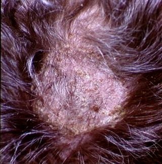
| 15:38, 3 March 2008 | Ringworm dog.jpg (file) |  |
79 KB | Bara | 1 | |
| 15:40, 3 March 2008 | Trichophyton mentagrophytes dog.jpg (file) |  |
268 KB | Bara | 1 | |
| 15:42, 3 March 2008 | Superficial pyoderma dog.jpg (file) |  |
69 KB | Bara | 1 | |
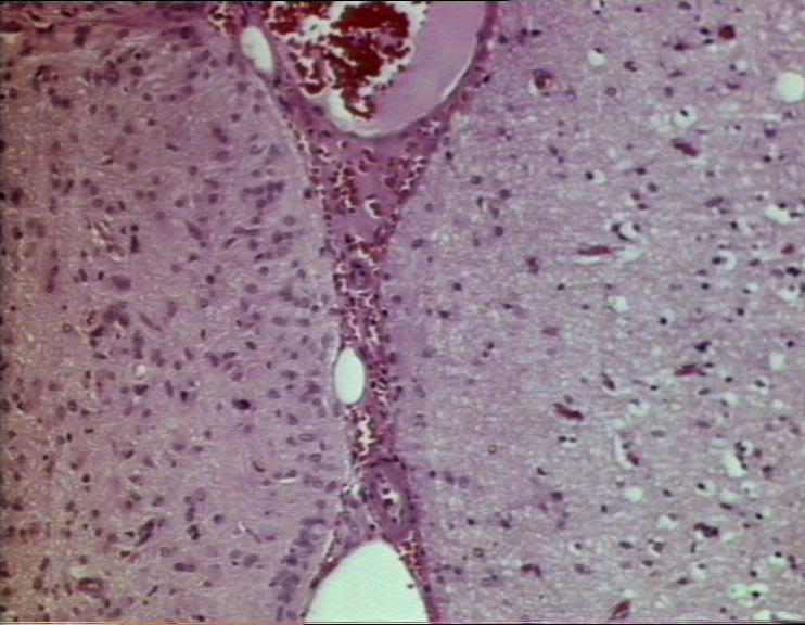
| 15:58, 25 March 2008 | Neuronalvacuolation1.jpg (file) |  |
59 KB | Lizzies | Neuronal vacuolation. Courtesy of BioMed Image Archive | 1 |
| 16:01, 25 March 2008 | Neuronalvacuolation2.jpg (file) |  |
59 KB | Lizzies | Neuronal Vacuolation. Courtesy of BioMed Image Archive. | 1 |
| 18:14, 25 March 2008 | Hydrocephalussection.jpg (file) |  |
58 KB | Lizzies | Section of a brain with internal hydrocephalus as a result of aqueduct stenosis. Courtesy of BioMed Image Archive. | 1 |
| 18:17, 25 March 2008 | Aqueductstenosis.jpg (file) |  |
74 KB | Lizzies | Aqueduct stenosis, causing hydrocephalus. Courtesy of BioMed Image Archive. | 1 |
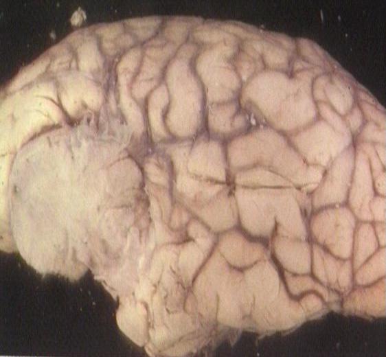
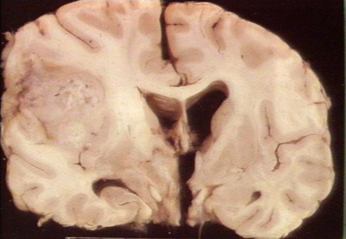
| 11:17, 26 March 2008 | Meningiomaleftfrontallobe.jpg (file) |  |
34 KB | Lizzies | Meningioma located at the left frontal lobe of the brain. Courtesy of BioMed Image Archive | 1 |
| 11:18, 26 March 2008 | Meningiomabrain.jpg (file) |  |
52 KB | Lizzies | Meningioma on the brain. Courtesy of BioMed Image Archive | 1 |
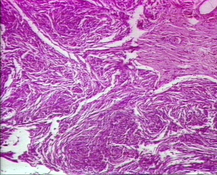
| 11:24, 26 March 2008 | Meningiomahisto.jpg (file) |  |
101 KB | Lizzies | Histological view of a meningioma. Courtesy of BioMed Image Archive. | 1 |
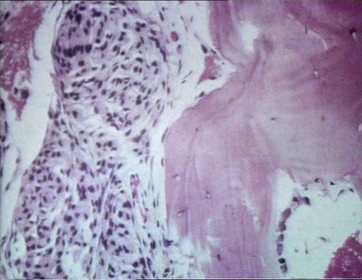
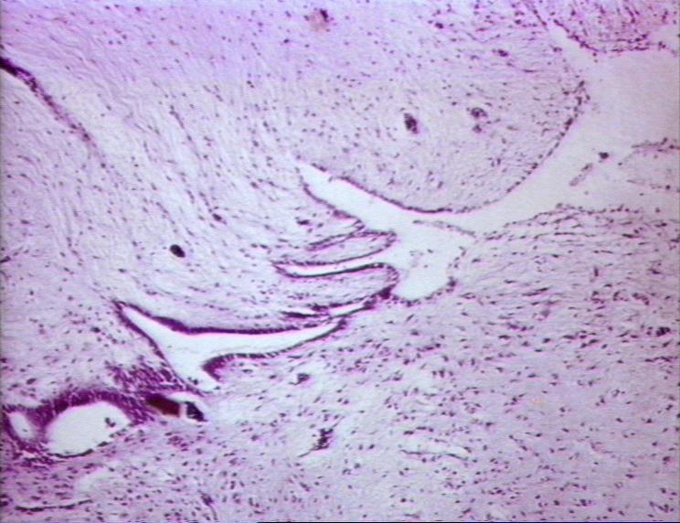
| 11:25, 26 March 2008 | Meningiomainfiltrating.jpg (file) |  |
56 KB | Lizzies | Meningioma infiltrating between bony trabeculae. Courtesy of BioMed Image Archive. | 1 |
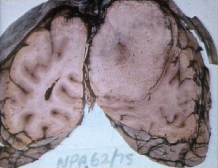
| 11:43, 26 March 2008 | Astrocytomagross.jpg (file) |  |
39 KB | Lizzies | Astrocytoma. Courtesy of BioMed Image Archive | 1 |
| 11:44, 26 March 2008 | Astrocytomahisto.jpg (file) |  |
68 KB | Lizzies | Astrocytoma - histological view. Courtesy of BioMed Image Archive. | 1 |
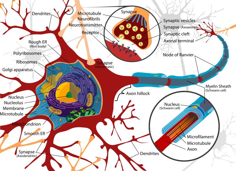
| 14:32, 26 March 2008 | Neurondiagram.jpg (file) |  |
101 KB | Lizzies | Diagram of a neuron. Image taken from WikiMedia Commons image repository. [http://commons.wikimedia.org/wiki/Image:Complete_neuron_cell_diagram.svg] | 1 |
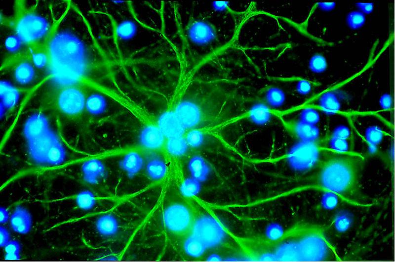
| 16:18, 26 March 2008 | Astrocyte.jpg (file) |  |
75 KB | Lizzies | An astrocyte in culture, stained immunofluorecently. The astrocyte processes are stained green, and the nuclei of this and other cells in the culture are stained blue. Imagae courtesy of [http://www2.unil.ch/edab/old/fr/presse_info.htm the Eurpoean Dana | 1 |
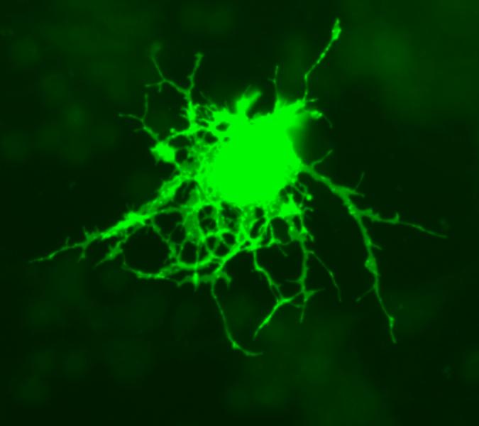
| 10:48, 27 March 2008 | Oligodendrocyte.jpg (file) |  |
22 KB | Lizzies | Oligodendrocyte. Image obtained from [http://commons.wikimedia.org/wiki/Image:Oligodendrocyte.png WikiMedia Commons]. | 1 |
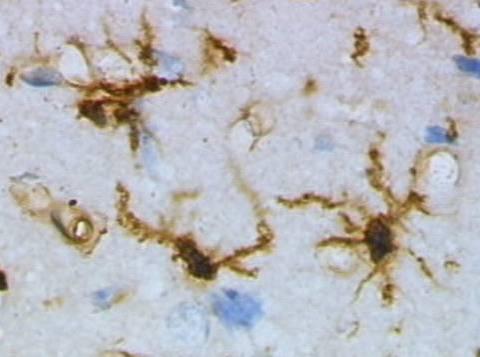
| 11:38, 27 March 2008 | Microglia.jpg (file) |  |
21 KB | Lizzies | Microglia cells stained immunohisotchemically for lectins. Image sourced from [http://commons.wikimedia.org/wiki/Image:Mikroglej_1.jpg|WikiMedia Commons], where it is attributed to Grzegorz Wicher. | 1 |
| 12:51, 27 March 2008 | Forkingaqueduct.jpg (file) |  |
73 KB | Lizzies | Forking aqueduct (an abnormal feature) lined with ependymal cells. Courtesy of BioMed Image Archive. | 1 |
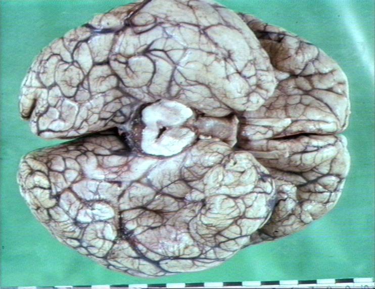
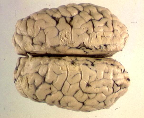
| 13:28, 27 March 2008 | Cerebralcortex.jpg (file) |  |
39 KB | Lizzies | Whole brain viewed from above showing cerebrum. Courtesy of BioMed Image Archive | 1 |
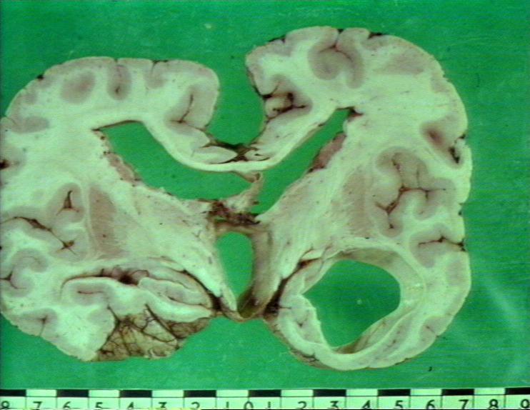
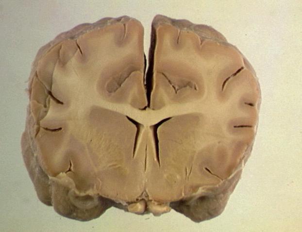
| 13:31, 27 March 2008 | Braincosssection.jpg (file) |  |
31 KB | Lizzies | Cross-section through the brain, showing the cerebrum, basal nuclei and lateral ventricle. The white and grey matter can be easily distinguished. | 1 |
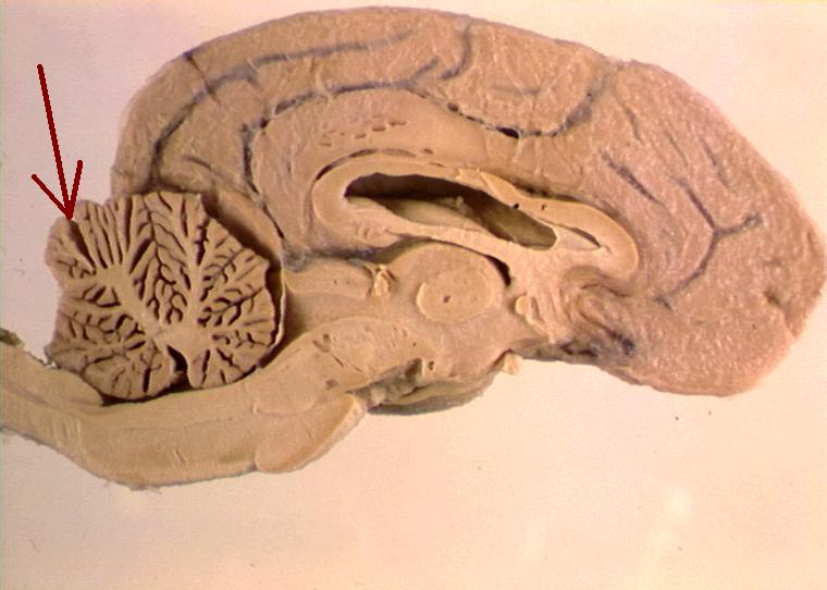
| 14:33, 27 March 2008 | Braincerebellumarrow.jpg (file) |  |
49 KB | Lizzies | Cross section of the brain. The arrow indicates the location of the cerebellum. Courtesy of BioMed Image Archive. | 1 |
| 08:09, 2 May 2008 | Ba 250 07.jpg (file) |  |
136 KB | Srhind | H&E ganglion | 1 |
| 13:02, 2 May 2008 | Thorax.jpg (file) |  |
57 KB | Srhind | Pleural effusion (hydrothorax) in dog with severe protein losing enteropathy | 1 |
| 13:10, 2 May 2008 | Coagulative necrosis.jpg (file) |  |
99 KB | Srhind | Coagulative necrosis in association with renal infarction | 1 |
| 07:17, 3 June 2008 | Congo red.jpg (file) |  |
98 KB | Srhind | Histopathology - amyloidosis. Stain: congo red | 1 |
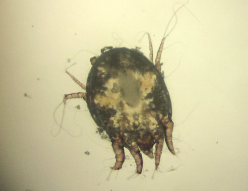
| 07:43, 19 June 2008 | Erythrophagocytosis.jpg (file) |  |
62 KB | Srhind | 1 | |
| 10:04, 19 June 2008 | Testclinpath.jpg (file) |  |
60 KB | Srhind | 1 | |
| 10:08, 19 June 2008 | Testclinpath150.jpg (file) |  |
71 KB | Srhind | 1 | |
| 10:09, 19 June 2008 | Neutstest.jpg (file) |  |
42 KB | Srhind | 1 | |
| 11:49, 19 June 2008 | REGANAEMIA.JPG (file) |  |
52 KB | Srhind | 1 | |
| 13:04, 19 June 2008 | REGANAEMIA.jpg (file) |  |
52 KB | Srhind | 1 | |
| 12:40, 22 June 2008 | Clinical Case 12 01.jpg (file) |  |
57 KB | Christina | 1 | |
| 12:56, 22 June 2008 | Clinical Case 12 02.jpg (file) |  |
28 KB | Christina | 1 | |
| 13:04, 22 June 2008 | Clinical Case 12 03.jpg (file) |  |
28 KB | Christina | 1 | |
| 13:07, 22 June 2008 | Clinical Case 12 04.jpg (file) |  |
31 KB | Christina | 1 | |
| 07:48, 23 June 2008 | Eosinophilic enteritis.jpg (file) |  |
142 KB | Srhind | Eosinophil rich inflammatory infiltrate in small intestine - muscularis layers | 1 |
| 10:10, 23 June 2008 | Lawsonia (1).jpg (file) | .jpg) |
141 KB | Srhind | H&E stained section small intestine. Pig 14 days post infection. | 1 |