File list
Jump to navigation
Jump to search
This special page shows all uploaded files.
| Date | Name | Thumbnail | Size | User | Description | Versions |
|---|---|---|---|---|---|---|
| 10:20, 11 October 2007 | Normal pancreas histo.jpg (file) |  |
100 KB | Bara | 1 | |
| 10:22, 11 October 2007 | Pancreatic carcinoma.jpg (file) |  |
50 KB | Bara | 1 | |
| 10:24, 11 October 2007 | Pancreatic carcinoma histo.jpg (file) |  |
66 KB | Bara | 1 | |
| 10:26, 11 October 2007 | Pancreatic atrophy.jpg (file) |  |
78 KB | Bara | 1 | |
| 10:30, 11 October 2007 | Pancreatic necrosis.jpg (file) |  |
60 KB | Bara | 1 | |
| 10:32, 11 October 2007 | Islet of Langerhans.jpg (file) |  |
100 KB | Bara | 1 | |
| 10:34, 11 October 2007 | Pancreatitis.jpg (file) |  |
81 KB | Bara | 1 | |
| 10:36, 11 October 2007 | Islet cell tumour.jpg (file) |  |
83 KB | Bara | 1 | |
| 01:55, 12 October 2007 | Anatomy picture.jpg (file) |  |
131 KB | Eabachynsky | 1 | |
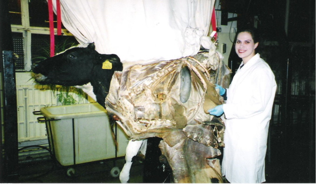
| 14:15, 3 December 2007 | Alimentary Case 1 Ultrasound.jpg (file) |  |
553 KB | Bara | 1 | |
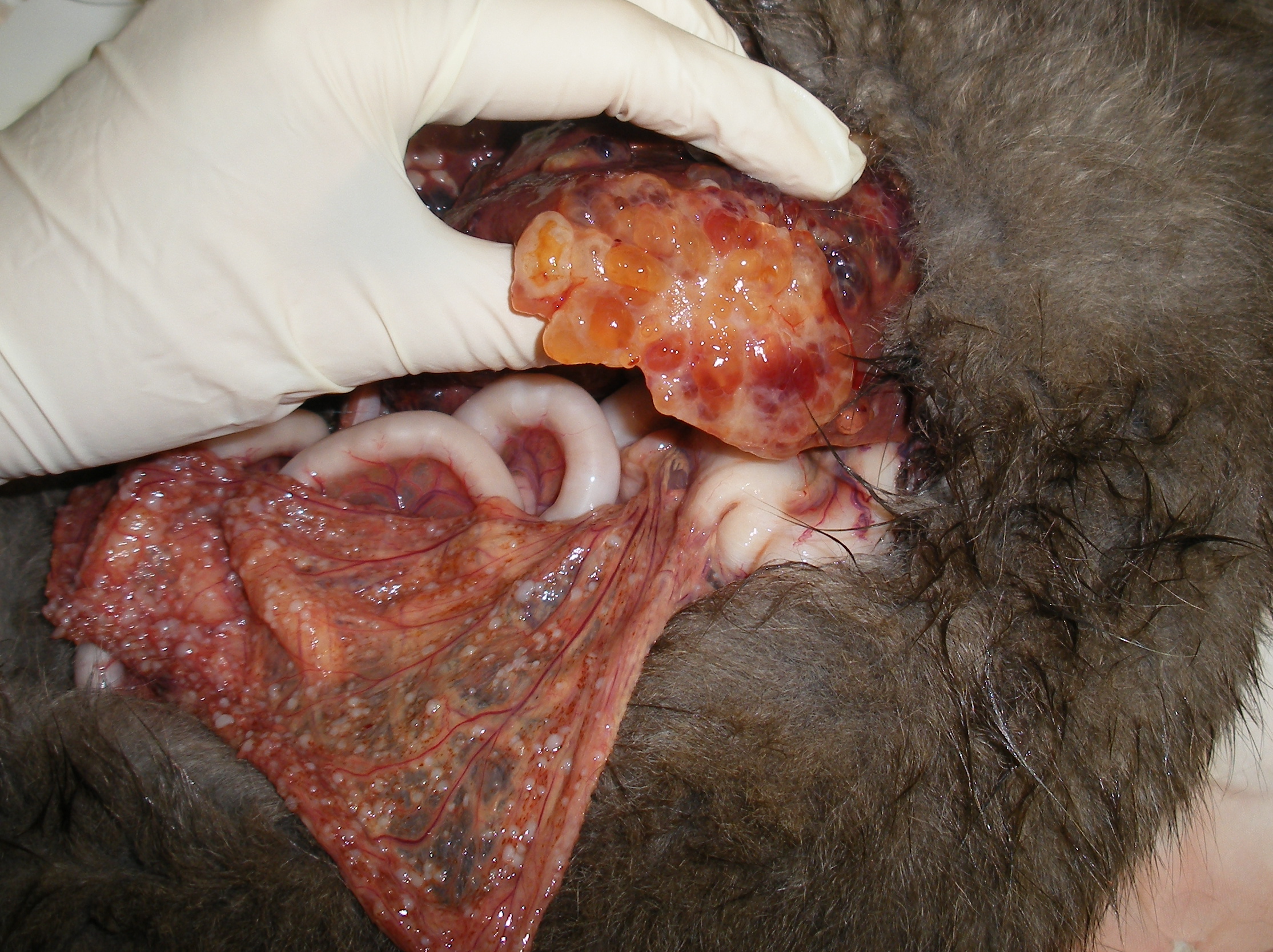
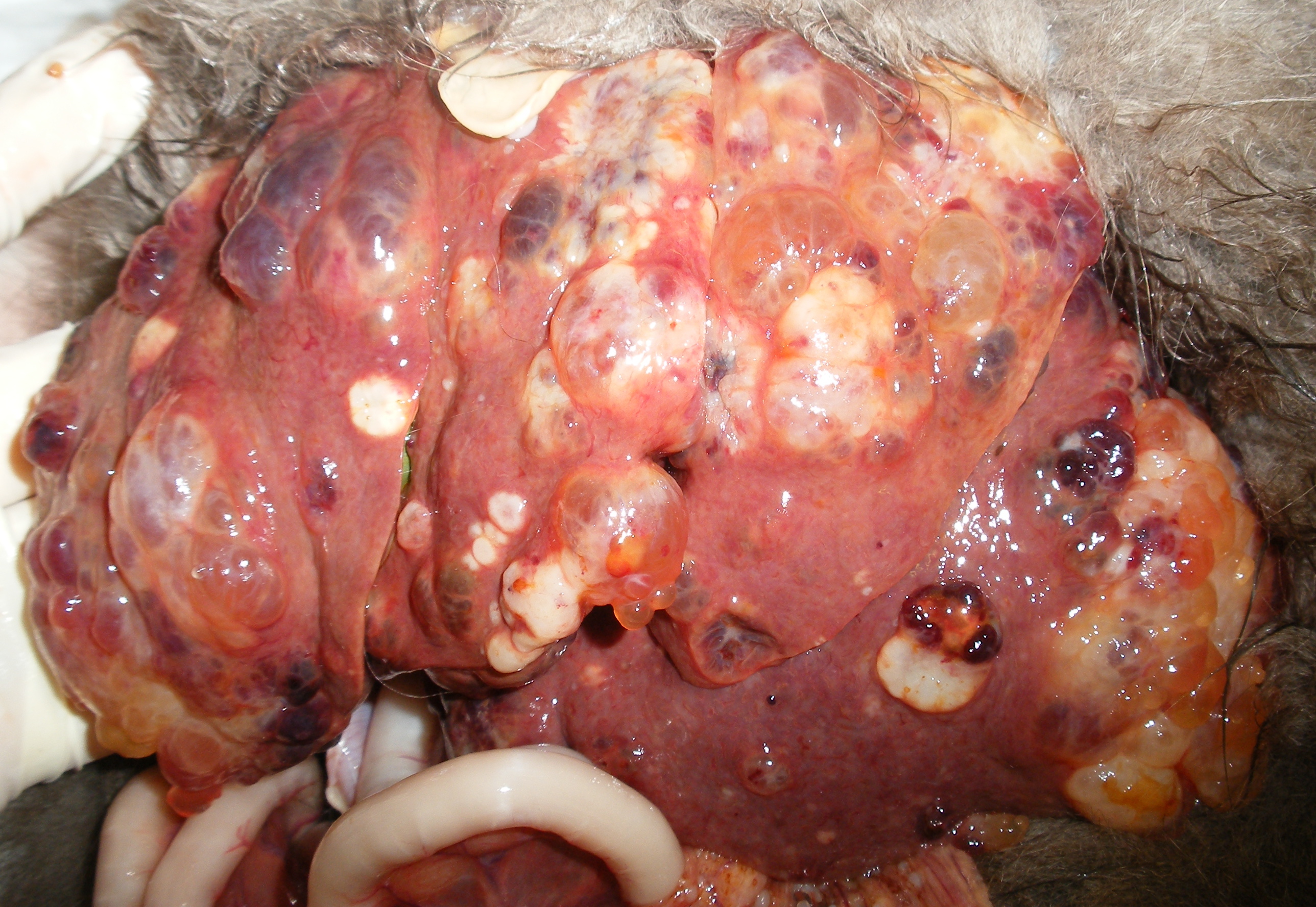
| 14:56, 3 December 2007 | Alimentary Case 1 liver 1.jpg (file) |  |
1.33 MB | Bara | 1 | |
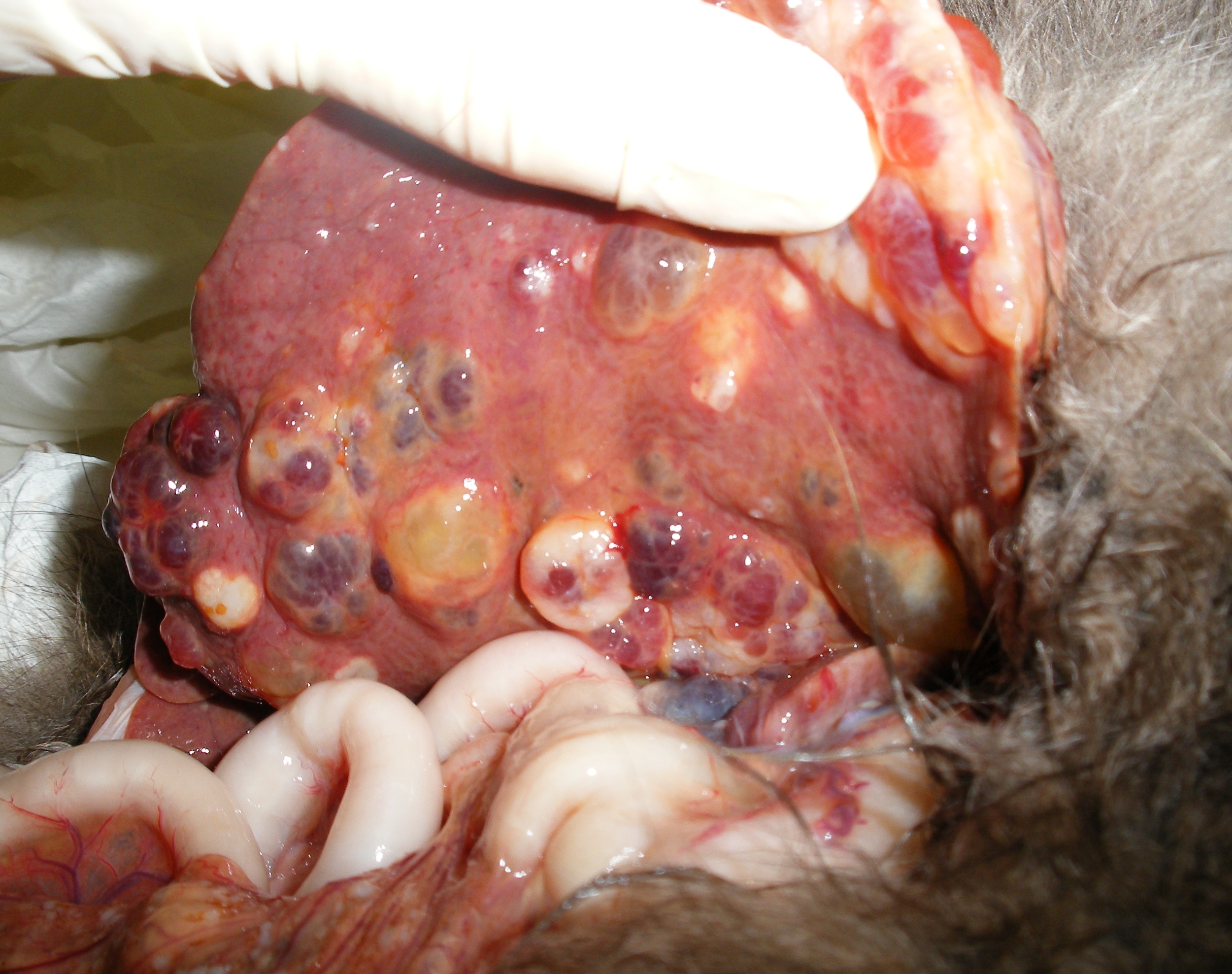
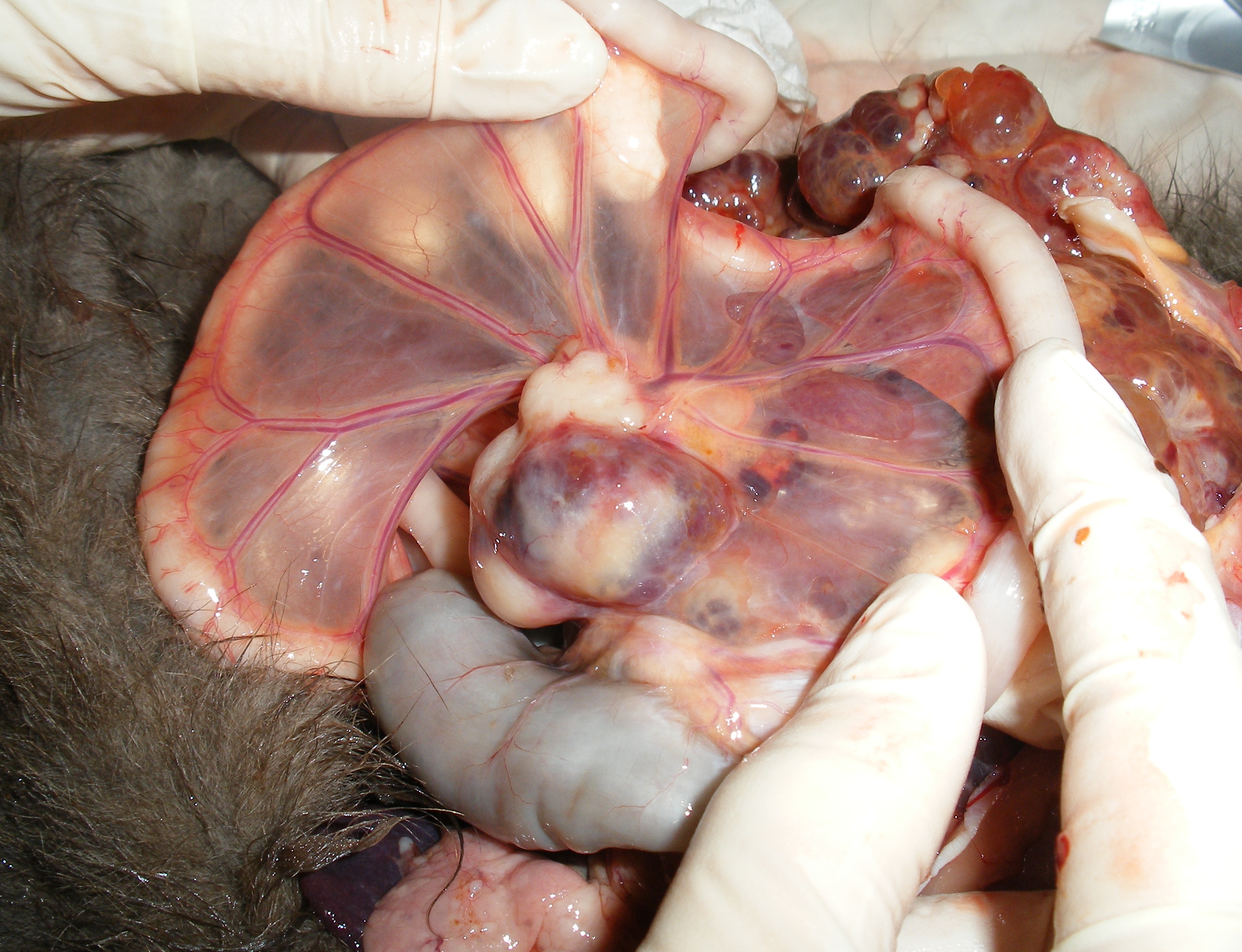
| 14:58, 3 December 2007 | Alimentary Case 1 liver 2.jpg (file) |  |
1.3 MB | Bara | Courtesy of A. Antonczyk | 1 |
| 15:00, 3 December 2007 | Alimentary Case 1 liver 3.jpg (file) |  |
1.5 MB | Bara | Courtesy of A. Antonczyk | 1 |
| 15:02, 3 December 2007 | Alimentary Case 1 liver 4.jpg (file) |  |
1.43 MB | Bara | Courtesy of A. Antonczyk | 1 |
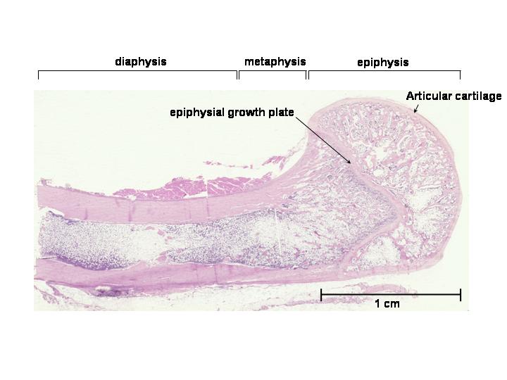
| 16:51, 3 December 2007 | Bone histo.jpg (file) |  |
39 KB | Bara | 1 | |
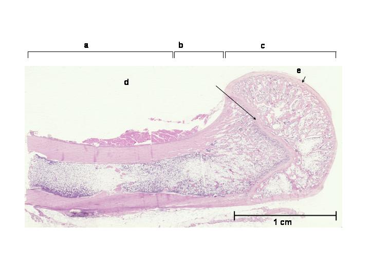
| 17:01, 3 December 2007 | Question bone histo.jpg (file) |  |
34 KB | Bara | 1 | |
| 16:06, 4 January 2008 | Normalparathyroid.jpg (file) |  |
71 KB | Lizzies | Normal histological appearance of the parathyroid gland. Courtesy of Biomed Image Archive. | 1 |
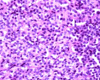
| 16:10, 4 January 2008 | Parathyroidadeoma.jpg (file) |  |
88 KB | Lizzies | Parathyroid adenoma, a cause of primary hyperparathyroidism. The image shows trabeculae of chief cells with lyaline stroma. Image courtesy of Biomed Image Archive. | 1 |
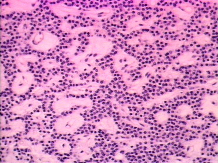
| 16:13, 4 January 2008 | Parathyroidhyperplasia.jpg (file) |  |
26 KB | Lizzies | Parathyroid gland hyperplasia, a cause of primary hyperparathyroidism. Image courtesy of Biomed Archive. | 1 |
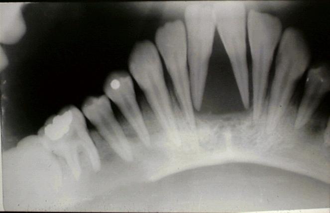
| 16:17, 4 January 2008 | Secondaryhyperparathyroidism.jpg (file) |  |
25 KB | Lizzies | Dental radiograph of secondary hyperparathyroidism. Note the demineralisation of bone around the tooth roots. Courtesy of Biomed Image Archive. | 1 |
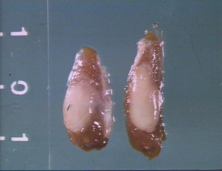
| 16:19, 4 January 2008 | Renalhyperparathyroidism.jpg (file) |  |
43 KB | Lizzies | Parathyroid hyperplasia seen in renal hyperparathyroidism. Courtesy of Biomed Image Archive. | 1 |
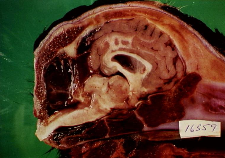
| 17:44, 4 January 2008 | Cerebellarhypoplasia.jpg (file) |  |
65 KB | Lizzies | Cross-section of head of seven-day-old calf showing cerebellar hypoplasia and atrophy of occipital cortical lobe. Courtesy of BioMed Image Archive. | 1 |
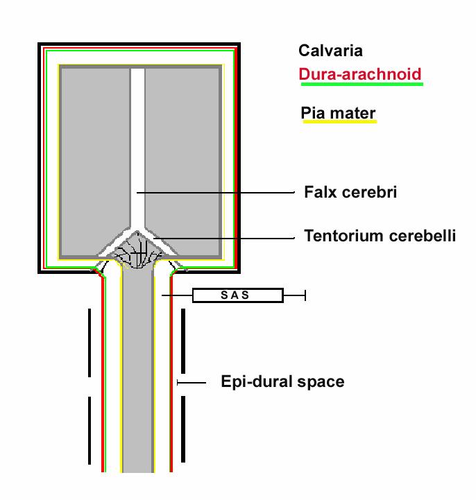
| 12:08, 10 January 2008 | Meningesdiagram.jpg (file) |  |
41 KB | Lizzies | The various layers enclosing the CNS. | 1 |
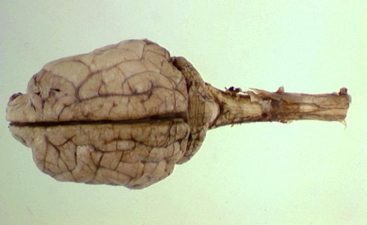
| 12:26, 10 January 2008 | Cerebrumbrainstemcerebellum.jpg (file) |  |
34 KB | Lizzies | Whole brain (canine) viewed from above showing the cerebrum, brain stem, and cerebellum. Courtesy of BioMed Image Archive | 1 |
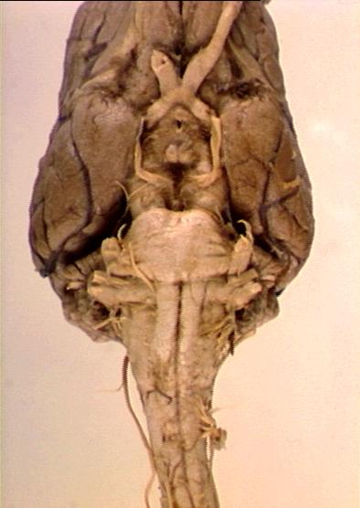
| 12:34, 10 January 2008 | Brainstemcranialnervespyramids.jpg (file) |  |
35 KB | Lizzies | Whole brain (canine) viewed from below showing brain stem, cranial nerves, and pyramids. Courtesy of BioMed Image Archive. | 1 |
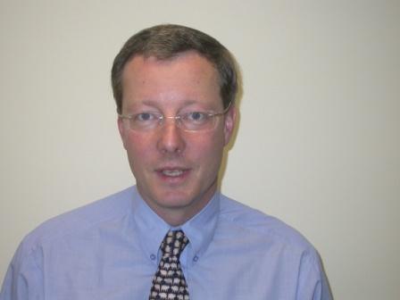
| 10:35, 16 January 2008 | MikeTargett.jpg (file) |  |
12 KB | MikeTargett | 1 | |
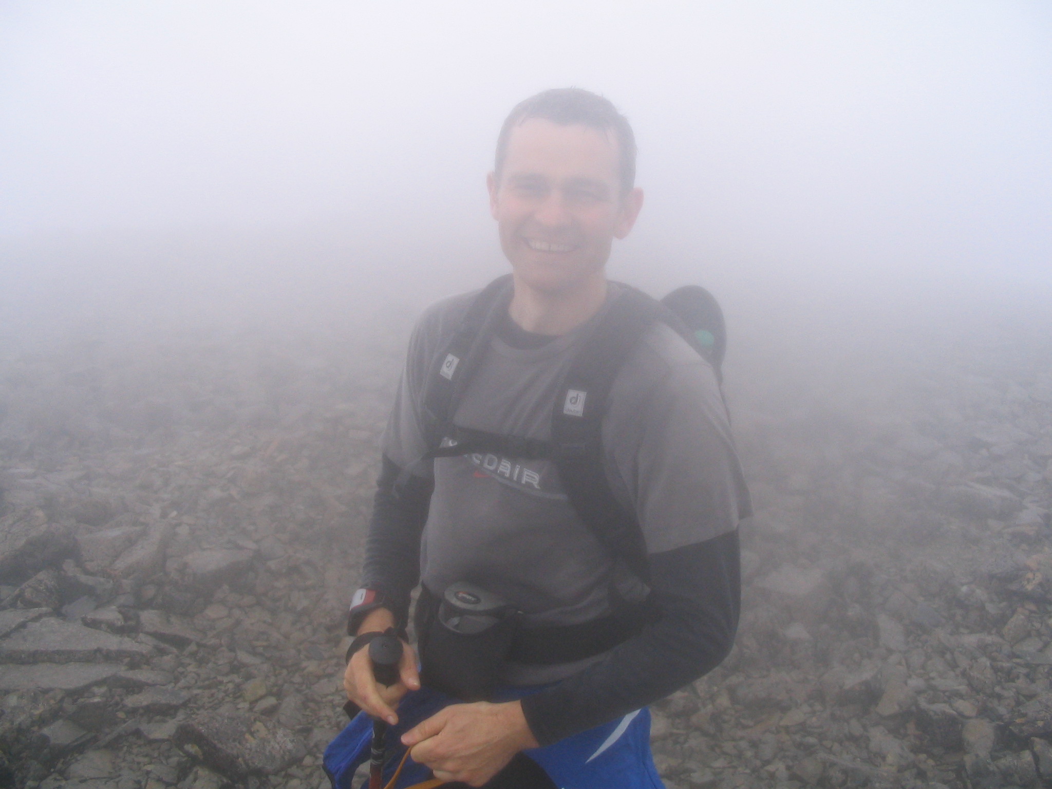
| 10:51, 16 January 2008 | IMG 1321.jpg (file) |  |
544 KB | Svzdg | 1 | |
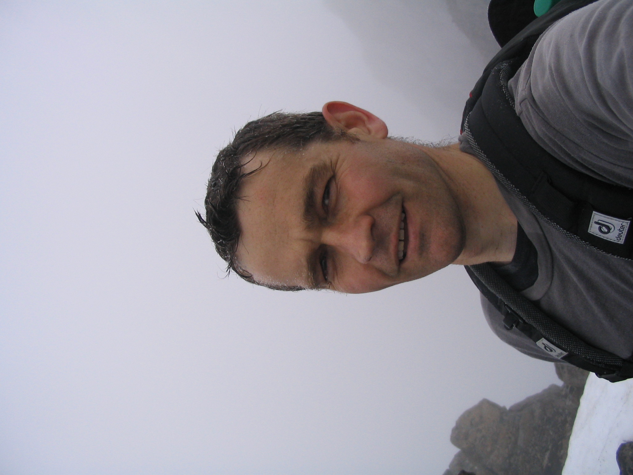
| 10:53, 16 January 2008 | Gardner David.JPG (file) |  |
510 KB | Svzdg | 1 | |
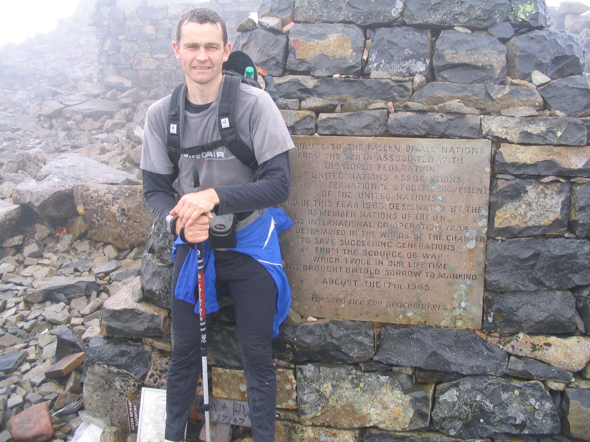
| 11:05, 16 January 2008 | IMG 1322.jpg (file) |  |
1.07 MB | Svzdg | Me at the top of Ben Nevis 2007, as part of a 3-peaks challenge | 1 |
| 15:49, 26 January 2008 | Clinical Case 2 01.jpg (file) |  |
133 KB | Christina | 1 | |
| 15:55, 26 January 2008 | Clinical Case 2 02.jpg (file) |  |
26 KB | Christina | 1 | |
| 15:56, 26 January 2008 | Clinical Case 2 03.jpg (file) |  |
34 KB | Christina | 1 | |
| 16:28, 26 January 2008 | Clinical Case 3 01.jpg (file) |  |
45 KB | Christina | 1 | |
| 16:29, 26 January 2008 | Clinical Case 3 02.jpg (file) |  |
46 KB | Christina | 1 | |
| 17:20, 26 January 2008 | Clinical Case 4 01.jpg (file) |  |
55 KB | Christina | 1 | |
| 17:49, 26 January 2008 | Clinical Case 4 02.jpg (file) |  |
36 KB | Christina | 1 | |
| 17:50, 26 January 2008 | Clinical Case 4 03.jpg (file) |  |
32 KB | Christina | 1 | |
| 17:41, 28 January 2008 | Whittlestone RCVS Blue Sky Application 23rd Jan Final.doc (file) | 202 KB | Kwhittlestone | Blue Sky Application 2008 | 1 | |
| 21:41, 28 January 2008 | Clinical Case 5 01.jpg (file) |  |
237 KB | Christina | 1 | |
| 21:43, 28 January 2008 | Clinical Case 5 02.jpg (file) |  |
237 KB | Christina | 1 | |
| 21:45, 28 January 2008 | Clinical Case 5 03.jpg (file) |  |
227 KB | Christina | 1 | |
| 19:43, 30 January 2008 | Clinical Case 6 01.jpg (file) |  |
83 KB | Christina | 1 | |
| 19:44, 30 January 2008 | Clinical Case 6 02.jpg (file) |  |
60 KB | Christina | 1 | |
| 19:52, 30 January 2008 | Clinical Case 6 04.jpg (file) |  |
36 KB | Christina | 1 | |
| 20:19, 30 January 2008 | Clinical Case 6 03.jpg (file) |  |
121 KB | Christina | 1 | |
| 12:49, 13 February 2008 | Pneumococcalmeningitis.jpg (file) |  |
52 KB | Lizzies | Pneumococcal meningitis. Courtesy of BioMed Image Archive. | 1 |
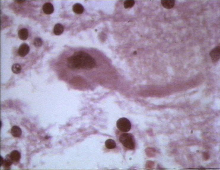
| 12:51, 13 February 2008 | Negribodies.jpg (file) |  |
53 KB | Lizzies | Negri bodies, seen in the neurons in rabies. Courtesy of BioMed Image Archive. | 1 |
| 20:15, 17 February 2008 | Clinical Case 7 01.jpg (file) |  |
45 KB | Christina | 1 | |
| 20:20, 17 February 2008 | Clinical Case 7 02.jpg (file) |  |
33 KB | Christina | 1 | |
| 20:23, 17 February 2008 | Clinical Case 7 03.jpg (file) |  |
24 KB | Christina | 1 |