File:SmAnOrth 01.jpg
Jump to navigation
Jump to search
No higher resolution available.
SmAnOrth_01.jpg (500 × 337 pixels, file size: 189 KB, MIME type: image/jpeg)
Summary
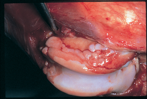
| Description |
Image from 'Small Animal Orthopaedics', with permission from Manson Publishing, as part of the OVAL. This is an intraoperative photograph of the trochlear ridges of the femoral condyle which have been exposed by a medial arthrotomy and lateral dislocation of the patella. |
|---|---|
| Date |
31/08/2011 |
| Source | |
| Author |
Daniel Lewis |
| Permission (Reusing this file) |
See below |
Licensing
| This file is licensed under the Creative Commons Attribution Non-Commercial & No Derivative Works License |
File history
Click on a date/time to view the file as it appeared at that time.
| Date/Time | Thumbnail | Dimensions | User | Comment | |
|---|---|---|---|---|---|
| current | 08:10, 2 September 2011 | | 500 × 337 (189 KB) | Ggaitskell (talk | contribs) | {{Information |Description =Image from [http://www.mansonpublishing.com/vet_titles/Lewis.html 'Small Animal Orthopaedics'], with permission from Manson Publishing, as part of the OVAL. This is an intraoperative photograph of the trochle |
You cannot overwrite this file.
File usage
The following page uses this file: