Unused files
The following files exist but are not embedded in any page. Please note that other web sites may link to a file with a direct URL, and so may still be listed here despite being in active use.
Showing below up to 50 results in range #111 to #160.
View (previous 50 | next 50) (20 | 50 | 100 | 250 | 500)
 Nocardiosis in a puma.jpeg 750 × 497; 310 KB
 Glasser's disease - severe acute fibrinous peritonitis.jpeg 750 × 493; 75 KB
 Acute peritonitis and cecal base rupture.jpeg 750 × 508; 100 KB
 Cysticercus pisiformis.jpeg 750 × 497; 276 KB
 Tubeculous peritonitis.jpeg 320 × 248; 13 KB
 Carcinomatosis and sclerosis in sheep.jpeg 750 × 499; 189 KB
 Lipoma in horse.jpeg 750 × 499; 63 KB
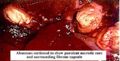
 Bovine pancreatic calculi.jpeg 750 × 594; 50 KB
 Pancreatic hypoplasia by King.jpeg 750 × 513; 50 KB
 Pancreatic adenoma cat.jpeg 750 × 499; 299 KB
 Ectopic pancreas.jpeg 1,796 × 1,076; 57 KB
 Insulinoma King.jpeg 750 × 498; 197 KB
 Pancreatic cysts by.jpeg 750 × 516; 352 KB
 Pancreatic flukes in a wolf by King.jpeg 750 × 500; 177 KB
 Fat necrosis by King.jpeg 750 × 487; 99 KB
 Pancreatic carcinoma.jpeg 750 × 480; 66 KB
 Strongylus equinus granulomas in pancreas.jpeg 750 × 515; 101 KB
 Gastrinoma by King.jpeg 750 × 496; 62 KB
 Peritoneal mesothelioma BioMed by King.jpeg 320 × 247; 16 KB
 Congenital umbilical hernia.jpeg 750 × 563; 53 KB
 Rupture uterus with fibrinous peritonitis in a cow.jpeg 750 × 504; 540 KB
 Rupture pyometra in a rabbit.jpeg 750 × 500; 173 KB
 Diaphragmatic hernia in a cat in RTA.jpeg 750 × 497; 64 KB
 Steatitis.jpeg 750 × 516; 62 KB
 Granulomatous fat necrosis in Guernsey.jpeg 750 × 514; 55 KB
 Cellular swelling diagram.jpg 710 × 543; 56 KB
 Hydropic degeneration foot and mouth pig foot.jpg 247 × 552; 23 KB
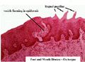
 Hydropic degneration foot and mouth ox tongue.jpg 292 × 391; 25 KB
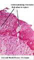
 Hydropic degeneration foot and mouth ox tongue histo 1.jpg 326 × 236; 16 KB
 Hydropic degeneration foot and mouth ox tongue histo 2.jpg 188 × 333; 17 KB
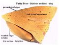 Fatty liver.jpg 404 × 306; 21 KB
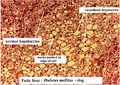 Fatty liver histo.jpg 547 × 387; 68 KB
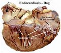 Endocardiosis.jpg 496 × 436; 45 KB
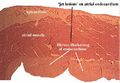 Endocardiosis histo 2.jpg 493 × 340; 35 KB
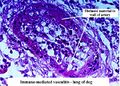 Fibrinoid degeneration immune mediated vasculitis.jpg 474 × 338; 54 KB
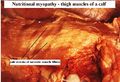 Nutritional myopathy.jpg 478 × 325; 34 KB
 Nutritional myopathy histo.jpg 453 × 318; 39 KB
 Amyloidosis.jpg 411 × 293; 29 KB
 Glycogen infiltration.jpg 496 × 350; 53 KB
 Viral inclusion canine adenovirus 1.jpg 363 × 237; 23 KB
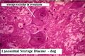 Lysosomal storage disease.jpg 526 × 347; 41 KB
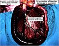 Strangulation of intestine.jpg 512 × 404; 49 KB
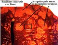 Coagulative necrosis bacillary necrosis.jpg 437 × 343; 35 KB
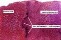 Coagulative necrosis histo.jpg 422 × 277; 35 KB
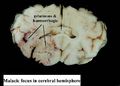 Malacia.jpg 448 × 320; 19 KB
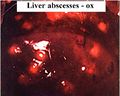 Liver abscess.jpg 379 × 304; 21 KB
 Abscess slice.jpg 506 × 240; 31 KB
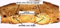
 Abscess centre and capsule.jpg 368 × 186; 19 KB
 Macrophage caseative necrosis.JPG 421 × 348; 39 KB


