File:Peribronchiolar Cuffing Enzootic Pneumonia1.jpg

Original file (767 × 612 pixels, file size: 123 KB, MIME type: image/jpeg)
Summary
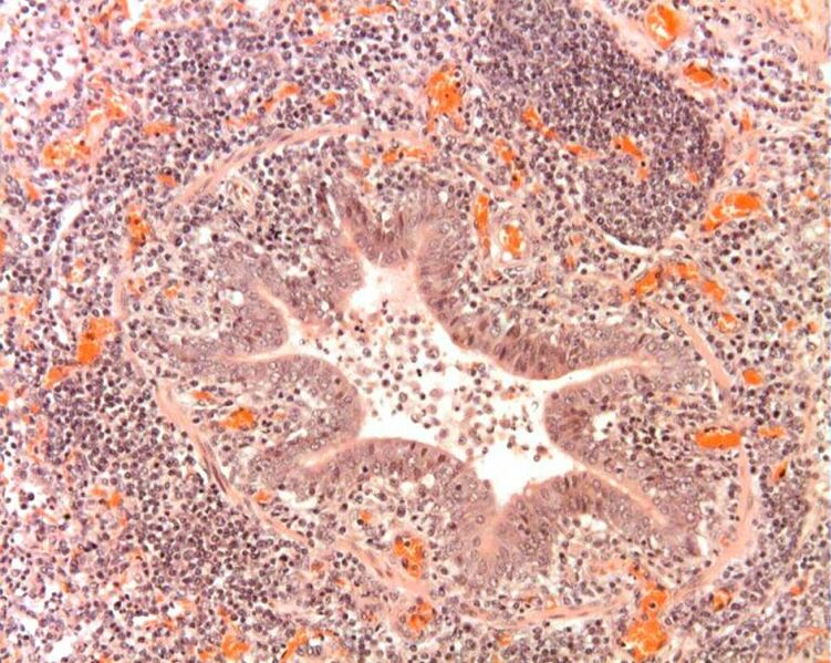
| Description |
Peribronchiolar cuffing in enzootic pneumonia. |
|---|---|
| Date |
uploaded September 2011 |
| Source |
Prof Andrew Rycroft |
| Author |
Andrew Rycroft |
| Permission (Reusing this file) |
See below |
Licensing
| This file is licensed under the Creative Commons Attribution Non-Commercial & No Derivative Works License |
File history
Click on a date/time to view the file as it appeared at that time.
| Date/Time | Thumbnail | Dimensions | User | Comment | |
|---|---|---|---|---|---|
| current | 17:21, 7 September 2011 | | 767 × 612 (123 KB) | HelenD (talk | contribs) | {{Information |Description=Peribronchiolar cuffing in enzootic pneumonia. |Source=Prof Andrew Rycroft |Date= |Author=Andrew Rycroft |Permission= |other_versions= }} |
You cannot overwrite this file.
File usage
The following page uses this file: